 Movie
Movie Controller

[English] 日本語
 Yorodumi
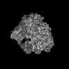
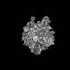
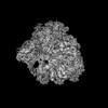
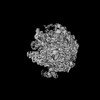
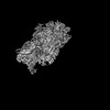
- PDB-8why: Cryo- EM structure of Mycobacterium smegmatis 50S ribosomal subun... -
+ Open data

- Basic information
| Entry | Database: PDB / ID: 8why | ||||||
|---|---|---|---|---|---|---|---|
| Title | Cryo- EM structure of Mycobacterium smegmatis 50S ribosomal subunit (body 1) of 70S ribosome and RafH. | ||||||
 Components Components |
| ||||||
 Keywords Keywords | RIBOSOME / protein synthesis / Mycobacterium smegmatis / hibernation promotion factor / RafH / hypoxia stress / Cryo- EM / Single particle reconstruction | ||||||
| Function / homology |  Function and homology information Function and homology informationlarge ribosomal subunit / transferase activity / 5S rRNA binding / ribosomal large subunit assembly / large ribosomal subunit rRNA binding / cytosolic large ribosomal subunit / cytoplasmic translation / tRNA binding / negative regulation of translation / rRNA binding ...large ribosomal subunit / transferase activity / 5S rRNA binding / ribosomal large subunit assembly / large ribosomal subunit rRNA binding / cytosolic large ribosomal subunit / cytoplasmic translation / tRNA binding / negative regulation of translation / rRNA binding / structural constituent of ribosome / ribosome / translation / ribonucleoprotein complex / mRNA binding / metal ion binding / cytoplasm Similarity search - Function | ||||||
| Biological species |  Mycolicibacterium smegmatis MC2 155 (bacteria) Mycolicibacterium smegmatis MC2 155 (bacteria) | ||||||
| Method | ELECTRON MICROSCOPY / single particle reconstruction / cryo EM / Resolution: 2.7 Å | ||||||
 Authors Authors | Kumar, N. / Sharma, S. / Kaushal, P.S. | ||||||
| Funding support |  India, 1items India, 1items
| ||||||
 Citation Citation | Journal: Nat Commun / Year: 2024 Title: Cryo- EM structure of the mycobacterial 70S ribosome in complex with ribosome hibernation promotion factor RafH. Authors: Niraj Kumar / Shivani Sharma / Prem S Kaushal / Abstract: Ribosome hibernation is a key survival strategy bacteria adopt under environmental stress, where a protein, hibernation promotion factor (HPF), transitorily inactivates the ribosome. Mycobacterium ...Ribosome hibernation is a key survival strategy bacteria adopt under environmental stress, where a protein, hibernation promotion factor (HPF), transitorily inactivates the ribosome. Mycobacterium tuberculosis encounters hypoxia (low oxygen) as a major stress in the host macrophages, and upregulates the expression of RafH protein, which is crucial for its survival. The RafH, a dual domain HPF, an orthologue of bacterial long HPF (HPF), hibernates ribosome in 70S monosome form, whereas in other bacteria, the HPF induces 70S ribosome dimerization and hibernates its ribosome in 100S disome form. Here, we report the cryo- EM structure of M. smegmatis, a close homolog of M. tuberculosis, 70S ribosome in complex with the RafH factor at an overall 2.8 Å resolution. The N- terminus domain (NTD) of RafH binds to the decoding center, similarly to HPF NTD. In contrast, the C- terminus domain (CTD) of RafH, which is larger than the HPF CTD, binds to a distinct site at the platform binding center of the ribosomal small subunit. The two domain-connecting linker regions, which remain mostly disordered in earlier reported HPF structures, interact mainly with the anti-Shine Dalgarno sequence of the 16S rRNA. | ||||||
| History |
|
- Structure visualization
| Structure viewer | Molecule: MolmilJmol/JSmol |
|---|
- Downloads & links
-Download
| PDBx/mmCIF format | 8why.cif.gz | 2.2 MB | Display | PDBx/mmCIF format |
|---|---|---|---|---|
| PDB format | pdb8why.ent.gz | 1.7 MB | Display | PDB format |
| PDBx/mmJSON format | 8why.json.gz | Tree view | PDBx/mmJSON format | |
| Others |  Other downloads Other downloads |
-Validation report
| Arichive directory | https://data.pdbj.org/pub/pdb/validation_reports/wh/8whyftp://data.pdbj.org/pub/pdb/validation_reports/wh/8why | HTTPS FTP |
|---|
-Related structure data
| Related structure data | 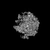 37552MC 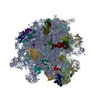 8whxC 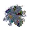 8wi7C  8wi8C 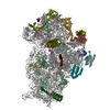 8wi9C 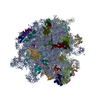 8wibC 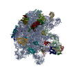 8wicC 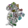 8widC 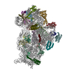 8wifC M: map data used to model this data C: citing same article ( |
|---|---|
| Similar structure data |


-Links
 PDBj
PDBj


- Assembly
| Deposited unit | 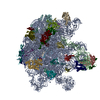
|
|---|---|
| 1 |
|
-Components
+50S ribosomal protein ... , 26 types, 26 molecules EFGHIJMNOQRSTUVWXZ12356784
-RNA chain , 2 types, 2 molecules AB
| #27: RNA chain | Mass: 1011834.750 Da / Num. of mol.: 1 / Source method: isolated from a natural source Source: (natural) Mycolicibacterium smegmatis MC2 155 (bacteria) |
|---|---|
| #28: RNA chain | Mass: 38061.816 Da / Num. of mol.: 1 / Source method: isolated from a natural source Source: (natural) Mycolicibacterium smegmatis MC2 155 (bacteria)Strain: mc(2)155 / References: GenBank: 118168627 |
-Experimental details
-Experiment
| Experiment | Method: ELECTRON MICROSCOPY |
|---|---|
| EM experiment | Aggregation state: PARTICLE / 3D reconstruction method: single particle reconstruction |
- Sample preparation
| Component | Name: 70S ribosome + RafH protein / Type: RIBOSOME / Entity ID: all / Source: NATURAL |
|---|---|
| Molecular weight | Experimental value: NO |
| Source (natural) | Organism: Mycolicibacterium smegmatis MC2 155 (bacteria) / Strain: mc(2)155 |
| Buffer solution | pH: 7.4 / Details: 20mM HEPES, 20mM MgCl2, 100mM NH4Cl, 3mM DTT |
| Specimen | Conc.: 1 mg/ml / Embedding applied: NO / Shadowing applied: NO / Staining applied: NO / Vitrification applied: YES |
| Specimen support | Grid material: COPPER / Grid mesh size: 300 divisions/in. / Grid type: Quantifoil R1.2/1.3 |
| Vitrification | Instrument: FEI VITROBOT MARK IV / Cryogen name: ETHANE / Humidity: 100 % |
- Electron microscopy imaging
| Experimental equipment |  Model: Titan Krios / Image courtesy: FEI Company |
|---|---|
| Microscopy | Model: FEI TITAN KRIOS |
| Electron gun | Electron source:  FIELD EMISSION GUN / Accelerating voltage: 300 kV / Illumination mode: OTHER FIELD EMISSION GUN / Accelerating voltage: 300 kV / Illumination mode: OTHER |
| Electron lens | Mode: BRIGHT FIELD / Nominal defocus max: 3000 nm / Nominal defocus min: 1800 nm |
| Specimen holder | Cryogen: NITROGEN / Specimen holder model: FEI TITAN KRIOS AUTOGRID HOLDER |
| Image recording | Average exposure time: 2 sec. / Electron dose: 1.34 e/Å2 / Detector mode: INTEGRATING / Film or detector model: FEI FALCON III (4k x 4k) / Num. of grids imaged: 3 / Num. of real images: 12343 |
- Processing
| EM software |
| |||||||||||||||||||||||||||||||||||||||||||||
|---|---|---|---|---|---|---|---|---|---|---|---|---|---|---|---|---|---|---|---|---|---|---|---|---|---|---|---|---|---|---|---|---|---|---|---|---|---|---|---|---|---|---|---|---|---|---|
| CTF correction | Details: CTF correction in Relion 3.1.4 / Type: NONE | |||||||||||||||||||||||||||||||||||||||||||||
| Particle selection | Num. of particles selected: 1202461 | |||||||||||||||||||||||||||||||||||||||||||||
| Symmetry | Point symmetry: C1 (asymmetric) | |||||||||||||||||||||||||||||||||||||||||||||
| 3D reconstruction | Resolution: 2.7 Å / Resolution method: FSC 0.143 CUT-OFF / Num. of particles: 110934 / Symmetry type: POINT | |||||||||||||||||||||||||||||||||||||||||||||
| Atomic model building | Protocol: FLEXIBLE FIT / Space: REAL / Details: phenix.real_space_refinement | |||||||||||||||||||||||||||||||||||||||||||||
| Atomic model building | PDB-ID: 8WHX Accession code: 8WHX / Source name: PDB / Type: experimental model | |||||||||||||||||||||||||||||||||||||||||||||
| Refine LS restraints |
|
