 Movie
Movie Controller

[English] 日本語
 Yorodumi
- PDB-8wd7: Structural organization of the palisade layer in isolated vaccini... -
+ Open data

- Basic information
| Entry | Database: PDB / ID: 8wd7 | ||||||
|---|---|---|---|---|---|---|---|
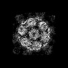
| Title | Structural organization of the palisade layer in isolated vaccinia virus cores | ||||||
 Components Components | Core protein OPG136 | ||||||
 Keywords Keywords | VIRAL PROTEIN / poxvirus / assembly / core / palisade layer / A10 | ||||||
| Function / homology | Poxvirus P4A / Poxvirus P4A protein / virion component / structural molecule activity / Major core protein OPG136 precursor Function and homology information Function and homology information | ||||||
| Biological species |  Vaccinia virus Vaccinia virus | ||||||
| Method | ELECTRON MICROSCOPY / subtomogram averaging / cryo EM / Resolution: 10 Å | ||||||
 Authors Authors | Liu, Y. / Qu, X. / Duan, M. / Shi, X. / Liu, S. / Shi, Y. / Gao, G.F. | ||||||
| Funding support |  China, 1items China, 1items
| ||||||
 Citation Citation | Journal: To Be Published Title: Cryo-ET reveals A10 protein as a major component of the poxvirus palisade layer Authors: Liu, Y. / Qu, X. / Duan, M. / Shi, X. / Liu, S. / Shi, Y. / Gao, G.F. | ||||||
| History |
|
- Structure visualization
| Structure viewer | Molecule: MolmilJmol/JSmol |
|---|
- Downloads & links
-Download
| PDBx/mmCIF format | 8wd7.cif.gz | 65.8 KB | Display | PDBx/mmCIF format |
|---|---|---|---|---|
| PDB format | pdb8wd7.ent.gz | 40.1 KB | Display | PDB format |
| PDBx/mmJSON format | 8wd7.json.gz | Tree view | PDBx/mmJSON format | |
| Others |  Other downloads Other downloads |
-Validation report
| Summary document | 8wd7_validation.pdf.gz | 1014.8 KB | Display | wwPDB validaton report |
|---|---|---|---|---|
| Full document | 8wd7_full_validation.pdf.gz | 1014.3 KB | Display | |
| Data in XML | 8wd7_validation.xml.gz | 28.3 KB | Display | |
| Data in CIF | 8wd7_validation.cif.gz | 43.9 KB | Display | |
| Arichive directory | https://data.pdbj.org/pub/pdb/validation_reports/wd/8wd7ftp://data.pdbj.org/pub/pdb/validation_reports/wd/8wd7 | HTTPS FTP |
-Related structure data
| Related structure data | 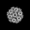 37456MC 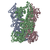 8wdcC M: map data used to model this data C: citing same article ( |
|---|---|
| Similar structure data |
-Links
 PDBj
PDBj- Assembly
| Deposited unit | 
|
|---|---|
| 1 |
|
-Components
| #1: Protein | Mass: 71073.477 Da / Num. of mol.: 3 / Source method: isolated from a natural source / Source: (natural) Vaccinia virus (strain Western Reserve) / Cell line: grown in HeLa cells / References: UniProt: P16715 |
|---|
-Experimental details
-Experiment
| Experiment | Method: ELECTRON MICROSCOPY |
|---|---|
| EM experiment | Aggregation state: PARTICLE / 3D reconstruction method: subtomogram averaging |
- Sample preparation
| Component | Name: Vaccinia virus Western Reserve / Type: VIRUS Details: Vaccinia virus was grown in HeLa cells and purified. The resultant mature virions were treated with DTT and NP40 and then purified to yield viral cores. Entity ID: all / Source: NATURAL |
|---|---|
| Molecular weight | Experimental value: NO |
| Source (natural) | Organism: Vaccinia virus Western Reserve |
| Details of virus | Empty: NO / Enveloped: YES / Isolate: STRAIN / Type: VIRION |
| Natural host | Organism: Homo sapiens |
| Buffer solution | pH: 9 / Details: 1mM Tris pH 9 |
| Specimen | Embedding applied: NO / Shadowing applied: NO / Staining applied: NO / Vitrification applied: YES Details: Vaccinia virus was grown in HeLa cells and purified. The resultant mature virions were treated with DTT and NP40 and then purified to yield viral cores. Isolated cores were used as a specimen. |
| Specimen support | Grid material: GOLD / Grid type: Quantifoil R2/2 |
| Vitrification | Instrument: FEI VITROBOT MARK IV / Cryogen name: ETHANE / Humidity: 100 % / Chamber temperature: 277 K |
- Electron microscopy imaging
| Experimental equipment |  Model: Titan Krios / Image courtesy: FEI Company |
|---|---|
| Microscopy | Model: FEI TITAN KRIOS |
| Electron gun | Electron source:  FIELD EMISSION GUN / Accelerating voltage: 300 kV / Illumination mode: FLOOD BEAM FIELD EMISSION GUN / Accelerating voltage: 300 kV / Illumination mode: FLOOD BEAM |
| Electron lens | Mode: BRIGHT FIELD / Nominal magnification: 53000 X / Nominal defocus max: 4500 nm / Nominal defocus min: 1600 nm / Cs: 0 mm / C2 aperture diameter: 70 µm / Alignment procedure: ZEMLIN TABLEAU |
| Specimen holder | Cryogen: NITROGEN / Specimen holder model: FEI TITAN KRIOS AUTOGRID HOLDER |
| Image recording | Electron dose: 2.9 e/Å2 / Avg electron dose per subtomogram: 119 e/Å2 / Film or detector model: FEI FALCON IV (4k x 4k) / Details: Dose rate 5.2 e-/pixel/s, EER (241 frames/sec) |
| EM imaging optics | Energyfilter name: TFS Selectris X Chromatic aberration corrector: Microscope was equipped with a Cc corrector Energyfilter slit width: 40 eV Spherical aberration corrector: Microscope was equipped with a Cs corrector |
- Processing
| EM software |
| ||||||||||||||||||||||||||||||||||||||||
|---|---|---|---|---|---|---|---|---|---|---|---|---|---|---|---|---|---|---|---|---|---|---|---|---|---|---|---|---|---|---|---|---|---|---|---|---|---|---|---|---|---|
| CTF correction | Type: PHASE FLIPPING AND AMPLITUDE CORRECTION | ||||||||||||||||||||||||||||||||||||||||
| Symmetry | Point symmetry: C3 (3 fold cyclic) | ||||||||||||||||||||||||||||||||||||||||
| 3D reconstruction | Resolution: 10 Å / Resolution method: FSC 0.143 CUT-OFF / Num. of particles: 207056 / Algorithm: FOURIER SPACE / Symmetry type: POINT | ||||||||||||||||||||||||||||||||||||||||
| EM volume selection | Details: Tomograms generated in Warp were denoised and corrected for missing wedge information using IsoNet. Subtomograms were selected by oversampling points at 4 nm distance using a surface model ...Details: Tomograms generated in Warp were denoised and corrected for missing wedge information using IsoNet. Subtomograms were selected by oversampling points at 4 nm distance using a surface model in Dynamo. In essence, for each viral core, a 3D surface was generated by fitting manually selected points on the core surface. Num. of tomograms: 74 / Num. of volumes extracted: 572775 | ||||||||||||||||||||||||||||||||||||||||
| Atomic model building | Source name: AlphaFold / Type: in silico model |
