[English] 日本語
 Yorodumi
- PDB-8wdc: Structural organization of the palisade layer in intracellular ma... -
+ Open data
- Basic information
| Entry | Database: PDB / ID: 8wdc | ||||||
|---|---|---|---|---|---|---|---|
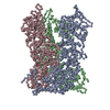
| Title | Structural organization of the palisade layer in intracellular mature vaccinia virions | ||||||
 Components Components | Core protein OPG136 | ||||||
 Keywords Keywords | VIRAL PROTEIN / poxvirus / assembly / core / palisade layer / A10 | ||||||
| Function / homology | Poxvirus P4A / Poxvirus P4A protein / virion component / structural molecule activity / Major core protein OPG136 precursor Function and homology information Function and homology information | ||||||
| Biological species |  Vaccinia virus Vaccinia virus | ||||||
| Method | ELECTRON MICROSCOPY / subtomogram averaging / cryo EM / Resolution: 7.7 Å | ||||||
 Authors Authors | Liu, Y. / Qu, X. / Duan, M. / Shi, X. / Liu, S. / Shi, Y. / Gao, G.F. | ||||||
| Funding support |  China, 1items China, 1items
| ||||||
 Citation Citation |  Journal: To Be Published Journal: To Be PublishedTitle: Cryo-ET reveals A10 protein as a major component of the poxvirus palisade layer Authors: Liu, Y. / Qu, X. / Duan, M. / Shi, X. / Liu, S. / Shi, Y. / Gao, G.F. | ||||||
| History |
|
- Structure visualization
| Structure viewer | Molecule:  Molmil Molmil Jmol/JSmol Jmol/JSmol |
|---|
- Downloads & links
- Download
| PDBx/mmCIF format |  8wdc.cif.gz 8wdc.cif.gz | 66.1 KB | Display |  PDBx/mmCIF format PDBx/mmCIF format |
|---|---|---|---|---|
| PDB format |  pdb8wdc.ent.gz pdb8wdc.ent.gz | 40.2 KB | Display |  PDB format PDB format |
| PDBx/mmJSON format |  8wdc.json.gz 8wdc.json.gz | Tree view |  PDBx/mmJSON format PDBx/mmJSON format | |
| Others |  Other downloads Other downloads |
-Validation report
| Summary document |  8wdc_validation.pdf.gz 8wdc_validation.pdf.gz | 1.1 MB | Display |  wwPDB validaton report wwPDB validaton report |
|---|---|---|---|---|
| Full document |  8wdc_full_validation.pdf.gz 8wdc_full_validation.pdf.gz | 1.1 MB | Display | |
| Data in XML |  8wdc_validation.xml.gz 8wdc_validation.xml.gz | 29.5 KB | Display | |
| Data in CIF |  8wdc_validation.cif.gz 8wdc_validation.cif.gz | 46.5 KB | Display | |
| Arichive directory |  https://data.pdbj.org/pub/pdb/validation_reports/wd/8wdc https://data.pdbj.org/pub/pdb/validation_reports/wd/8wdc ftp://data.pdbj.org/pub/pdb/validation_reports/wd/8wdc ftp://data.pdbj.org/pub/pdb/validation_reports/wd/8wdc | HTTPS FTP |
-Related structure data
| Related structure data |  37461MC  8wd7C M: map data used to model this data C: citing same article ( |
|---|---|
| Similar structure data | Similarity search - Function & homology  F&H Search F&H Search |
- Links
Links
- Assembly
| Deposited unit | 
|
|---|---|
| 1 |
|
- Components
| #1: Protein | Mass: 71073.477 Da / Num. of mol.: 3 / Source method: isolated from a natural source / Source: (natural)  Vaccinia virus (strain Western Reserve) / Cell line: grown in HeLa cells / References: UniProt: P16715 Vaccinia virus (strain Western Reserve) / Cell line: grown in HeLa cells / References: UniProt: P16715 |
|---|
-Experimental details
-Experiment
| Experiment | Method: ELECTRON MICROSCOPY |
|---|---|
| EM experiment | Aggregation state: PARTICLE / 3D reconstruction method: subtomogram averaging |
- Sample preparation
| Component | Name: Vaccinia virus Western Reserve / Type: VIRUS Details: Vaccinia virus was grown in HeLa cells and purified. Entity ID: all / Source: NATURAL |
|---|---|
| Molecular weight | Experimental value: NO |
| Source (natural) | Organism:  Vaccinia virus Western Reserve Vaccinia virus Western Reserve |
| Details of virus | Empty: NO / Enveloped: YES / Isolate: STRAIN / Type: VIRION |
| Natural host | Organism: Homo sapiens |
| Buffer solution | pH: 9 / Details: 1mM Tris pH 9 |
| Specimen | Embedding applied: NO / Shadowing applied: NO / Staining applied: NO / Vitrification applied: YES Details: Vaccinia virus was grown in HeLa cells and purified. The resultant mature virions were used as a specimen. |
| Specimen support | Grid material: GOLD / Grid type: Quantifoil R2/2 |
| Vitrification | Instrument: FEI VITROBOT MARK IV / Cryogen name: ETHANE / Humidity: 100 % / Chamber temperature: 277 K |
- Electron microscopy imaging
| Experimental equipment |  Model: Titan Krios / Image courtesy: FEI Company |
|---|---|
| Microscopy | Model: FEI TITAN KRIOS |
| Electron gun | Electron source:  FIELD EMISSION GUN / Accelerating voltage: 300 kV / Illumination mode: FLOOD BEAM FIELD EMISSION GUN / Accelerating voltage: 300 kV / Illumination mode: FLOOD BEAM |
| Electron lens | Mode: BRIGHT FIELD / Nominal magnification: 53000 X / Nominal defocus max: 3700 nm / Nominal defocus min: 1600 nm / Cs: 0 mm / C2 aperture diameter: 70 µm / Alignment procedure: ZEMLIN TABLEAU |
| Specimen holder | Cryogen: NITROGEN / Specimen holder model: FEI TITAN KRIOS AUTOGRID HOLDER |
| Image recording | Electron dose: 2.9 e/Å2 / Avg electron dose per subtomogram: 119 e/Å2 / Film or detector model: FEI FALCON IV (4k x 4k) / Details: Dose rate 5.2 e-/pixel/s, EER (241 frames/sec) |
| EM imaging optics | Energyfilter name: TFS Selectris X Chromatic aberration corrector: Microscope was equipped with a Cc corrector Details: about 20% of tilt series were collected using a slit width of 60 ev Energyfilter slit width: 40 eV Spherical aberration corrector: Microscope was equipped with a Cs corrector |
- Processing
| EM software |
| ||||||||||||||||||||||||||||||||||||||||
|---|---|---|---|---|---|---|---|---|---|---|---|---|---|---|---|---|---|---|---|---|---|---|---|---|---|---|---|---|---|---|---|---|---|---|---|---|---|---|---|---|---|
| CTF correction | Type: PHASE FLIPPING AND AMPLITUDE CORRECTION | ||||||||||||||||||||||||||||||||||||||||
| Symmetry | Point symmetry: C3 (3 fold cyclic) | ||||||||||||||||||||||||||||||||||||||||
| 3D reconstruction | Resolution: 7.7 Å / Resolution method: FSC 0.143 CUT-OFF / Num. of particles: 602843 / Algorithm: FOURIER SPACE / Symmetry type: POINT | ||||||||||||||||||||||||||||||||||||||||
| EM volume selection | Details: Tomograms generated in Warp were denoised and corrected for missing wedge information using IsoNet. Subtomograms were selected by oversampling points at 4 nm distance using a surface model ...Details: Tomograms generated in Warp were denoised and corrected for missing wedge information using IsoNet. Subtomograms were selected by oversampling points at 4 nm distance using a surface model in Dynamo. In essence, for each viral core, a 3D surface was generated by fitting manually selected points on the core surface. Num. of tomograms: 197 / Num. of volumes extracted: 1258156 | ||||||||||||||||||||||||||||||||||||||||
| Atomic model building | Source name: AlphaFold / Type: in silico model |
 Movie
Movie Controller


 PDBj
PDBj