[English] 日本語
 Yorodumi
- PDB-8wbg: CryoEM structure of non-structural protein 1 tetramer from ZIKA virus -
+ Open data
- Basic information
| Entry | Database: PDB / ID: 8wbg | ||||||
|---|---|---|---|---|---|---|---|
| Title | CryoEM structure of non-structural protein 1 tetramer from ZIKA virus | ||||||
 Components Components | Genome polyprotein | ||||||
 Keywords Keywords | VIRAL PROTEIN / flavivirus / non-structural protein 1 | ||||||
| Function / homology |  Function and homology information Function and homology informationsymbiont-mediated suppression of host JAK-STAT cascade via inhibition of STAT2 activity / ribonucleoside triphosphate phosphatase activity / double-stranded RNA binding / viral capsid / mRNA (nucleoside-2'-O-)-methyltransferase activity / mRNA 5'-cap (guanine-N7-)-methyltransferase activity / RNA helicase activity / protein dimerization activity / host cell endoplasmic reticulum membrane / symbiont-mediated suppression of host type I interferon-mediated signaling pathway ...symbiont-mediated suppression of host JAK-STAT cascade via inhibition of STAT2 activity / ribonucleoside triphosphate phosphatase activity / double-stranded RNA binding / viral capsid / mRNA (nucleoside-2'-O-)-methyltransferase activity / mRNA 5'-cap (guanine-N7-)-methyltransferase activity / RNA helicase activity / protein dimerization activity / host cell endoplasmic reticulum membrane / symbiont-mediated suppression of host type I interferon-mediated signaling pathway / symbiont-mediated suppression of host innate immune response / symbiont entry into host cell / viral RNA genome replication / serine-type endopeptidase activity / RNA-dependent RNA polymerase activity / virus-mediated perturbation of host defense response / fusion of virus membrane with host endosome membrane / host cell nucleus / virion attachment to host cell / structural molecule activity / virion membrane / proteolysis / extracellular region / ATP binding / membrane / metal ion binding Similarity search - Function | ||||||
| Biological species |  dengue virus type 4 dengue virus type 4 | ||||||
| Method | ELECTRON MICROSCOPY / single particle reconstruction / cryo EM / Resolution: 3.2 Å | ||||||
 Authors Authors | Jiao, H.Z. / Pan, Q. / Hu, H.L. | ||||||
| Funding support | 1items
| ||||||
 Citation Citation |  Journal: Sci Adv / Year: 2024 Journal: Sci Adv / Year: 2024Title: The step-by-step assembly mechanism of secreted flavivirus NS1 tetramer and hexamer captured at atomic resolution. Authors: Qi Pan / Haizhan Jiao / Wanqin Zhang / Qiang Chen / Geshu Zhang / Jianhai Yu / Wei Zhao / Hongli Hu /  Abstract: Flaviviruses encode a conserved, membrane-associated nonstructural protein 1 (NS1) with replication and immune evasion functions. The current knowledge of secreted NS1 (sNS1) oligomers is based on ...Flaviviruses encode a conserved, membrane-associated nonstructural protein 1 (NS1) with replication and immune evasion functions. The current knowledge of secreted NS1 (sNS1) oligomers is based on several low-resolution structures, thus hindering the development of drugs and vaccines against flaviviruses. Here, we revealed that recombinant sNS1 from flaviviruses exists in a dynamic equilibrium of dimer-tetramer-hexamer states. Two DENV4 hexameric NS1 structures and several tetrameric NS1 structures from multiple flaviviruses were solved at atomic resolution by cryo-EM. The stacking of the tetrameric NS1 and hexameric NS1 is facilitated by the hydrophobic β-roll and connector domains. Additionally, a triacylglycerol molecule located within the central cavity may play a role in stabilizing the hexamer. Based on differentiated interactions between the dimeric NS1, two distinct hexamer models (head-to-head and side-to-side hexamer) and the step-by-step assembly mechanisms of NS1 dimer into hexamer were proposed. We believe that our study sheds light on the understanding of the NS1 oligomerization and contributes to NS1-based therapies. | ||||||
| History |
|
- Structure visualization
| Structure viewer | Molecule:  Molmil Molmil Jmol/JSmol Jmol/JSmol |
|---|
- Downloads & links
- Download
| PDBx/mmCIF format |  8wbg.cif.gz 8wbg.cif.gz | 273.6 KB | Display |  PDBx/mmCIF format PDBx/mmCIF format |
|---|---|---|---|---|
| PDB format |  pdb8wbg.ent.gz pdb8wbg.ent.gz | 221.1 KB | Display |  PDB format PDB format |
| PDBx/mmJSON format |  8wbg.json.gz 8wbg.json.gz | Tree view |  PDBx/mmJSON format PDBx/mmJSON format | |
| Others |  Other downloads Other downloads |
-Validation report
| Summary document |  8wbg_validation.pdf.gz 8wbg_validation.pdf.gz | 1.5 MB | Display |  wwPDB validaton report wwPDB validaton report |
|---|---|---|---|---|
| Full document |  8wbg_full_validation.pdf.gz 8wbg_full_validation.pdf.gz | 1.5 MB | Display | |
| Data in XML |  8wbg_validation.xml.gz 8wbg_validation.xml.gz | 48.5 KB | Display | |
| Data in CIF |  8wbg_validation.cif.gz 8wbg_validation.cif.gz | 72.2 KB | Display | |
| Arichive directory |  https://data.pdbj.org/pub/pdb/validation_reports/wb/8wbg https://data.pdbj.org/pub/pdb/validation_reports/wb/8wbg ftp://data.pdbj.org/pub/pdb/validation_reports/wb/8wbg ftp://data.pdbj.org/pub/pdb/validation_reports/wb/8wbg | HTTPS FTP |
-Related structure data
| Related structure data |  37424MC 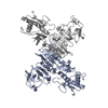 8wbbC 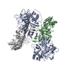 8wbcC 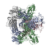 8wbdC 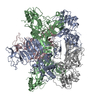 8wbeC 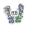 8wbfC 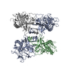 8wbhC M: map data used to model this data C: citing same article ( |
|---|---|
| Similar structure data | Similarity search - Function & homology  F&H Search F&H Search |
- Links
Links
- Assembly
| Deposited unit | 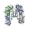
|
|---|---|
| 1 |
|
- Components
| #1: Protein | Mass: 44498.160 Da / Num. of mol.: 4 Source method: isolated from a genetically manipulated source Source: (gene. exp.)  dengue virus type 4 / Production host: dengue virus type 4 / Production host:  |
|---|
-Experimental details
-Experiment
| Experiment | Method: ELECTRON MICROSCOPY |
|---|---|
| EM experiment | Aggregation state: PARTICLE / 3D reconstruction method: single particle reconstruction |
- Sample preparation
| Component | Name: ZIKA virus non-structural protein 1 tetramer / Type: COMPLEX / Entity ID: all / Source: RECOMBINANT |
|---|---|
| Source (natural) | Organism:  dengue virus type 4 dengue virus type 4 |
| Source (recombinant) | Organism:  Homo sapiens (human) Homo sapiens (human) |
| Buffer solution | pH: 7.5 |
| Specimen | Embedding applied: NO / Shadowing applied: NO / Staining applied: NO / Vitrification applied: YES |
| Vitrification | Cryogen name: ETHANE / Humidity: 100 % |
- Electron microscopy imaging
| Experimental equipment |  Model: Titan Krios / Image courtesy: FEI Company |
|---|---|
| Microscopy | Model: FEI TITAN KRIOS |
| Electron gun | Electron source:  FIELD EMISSION GUN / Accelerating voltage: 300 kV / Illumination mode: FLOOD BEAM FIELD EMISSION GUN / Accelerating voltage: 300 kV / Illumination mode: FLOOD BEAM |
| Electron lens | Mode: BRIGHT FIELD / Nominal magnification: 105000 X / Nominal defocus max: 1800 nm / Nominal defocus min: 1200 nm / Cs: 2.7 mm / C2 aperture diameter: 100 µm |
| Image recording | Electron dose: 54 e/Å2 / Film or detector model: GATAN K3 BIOQUANTUM (6k x 4k) |
- Processing
| CTF correction | Type: PHASE FLIPPING AND AMPLITUDE CORRECTION |
|---|---|
| 3D reconstruction | Resolution: 3.2 Å / Resolution method: FSC 0.143 CUT-OFF / Num. of particles: 127526 / Symmetry type: POINT |
 Movie
Movie Controller


 PDBj
PDBj

