 Movie
Movie Controller


+ Open data

- Basic information
| Entry | Database: PDB / ID: 8w9r | |||||||||||||||||||||
|---|---|---|---|---|---|---|---|---|---|---|---|---|---|---|---|---|---|---|---|---|---|---|
| Title | Structure of Banna virus core | |||||||||||||||||||||
 Components Components |
| |||||||||||||||||||||
 Keywords Keywords | VIRUS / core particle / capsid protein / reovirus / complex | |||||||||||||||||||||
| Function / homology | Structural protein Vp10, Seadornavirus / Seadornavirus VP8 protein / Seadornavirus Vp10 / Seadornavirus outer capsid protein VP8 / Seadornavirus VP2 protein / Seadornavirus inner capsid protein VP2 / VP10 / Vp2 / VP8 Function and homology information Function and homology information | |||||||||||||||||||||
| Biological species |  Banna virus Banna virus | |||||||||||||||||||||
| Method | ELECTRON MICROSCOPY / single particle reconstruction / cryo EM / Resolution: 4.8 Å | |||||||||||||||||||||
 Authors Authors | Li, Z. / Cao, S. | |||||||||||||||||||||
| Funding support |  China, 1items China, 1items
| |||||||||||||||||||||
 Citation Citation | Journal: Nat Commun / Year: 2024 Title: Cryo-EM structures of Banna virus in multiple states reveal stepwise detachment of viral spikes. Authors: Zhiqiang Li / Han Xia / Guibo Rao / Yan Fu / Tingting Chong / Kexing Tian / Zhiming Yuan / Sheng Cao / Abstract: Banna virus (BAV) is the prototype Seadornavirus, a class of reoviruses for which there has been little structural study. Here, we report atomic cryo-EM structures of three states of BAV virions- ...Banna virus (BAV) is the prototype Seadornavirus, a class of reoviruses for which there has been little structural study. Here, we report atomic cryo-EM structures of three states of BAV virions-surrounded by 120 spikes (full virions), 60 spikes (partial virions), or no spikes (cores). BAV cores are double-layered particles similar to the cores of other non-turreted reoviruses, except for an additional protein component in the outer capsid shell, VP10. VP10 was identified to be a cementing protein that plays a pivotal role in the assembly of BAV virions by directly interacting with VP2 (inner capsid), VP8 (outer capsid), and VP4 (spike). Viral spikes (VP4/VP9 heterohexamers) are situated on top of VP10 molecules in full or partial virions. Asymmetrical electrostatic interactions between VP10 monomers and VP4 trimers are disrupted by high pH treatment, which is thus a simple way to produce BAV cores. Low pH treatment of BAV virions removes only the flexible receptor binding protein VP9 and triggers significant conformational changes in the membrane penetration protein VP4. BAV virions adopt distinct spatial organization of their surface proteins compared with other well-studied reoviruses, suggesting that BAV may have a unique mechanism of penetration of cellular endomembranes. | |||||||||||||||||||||
| History |
|
- Structure visualization
| Structure viewer | Molecule: MolmilJmol/JSmol |
|---|
- Downloads & links
-Download
| PDBx/mmCIF format | 8w9r.cif.gz | 1.1 MB | Display | PDBx/mmCIF format |
|---|---|---|---|---|
| PDB format | pdb8w9r.ent.gz | 934.5 KB | Display | PDB format |
| PDBx/mmJSON format | 8w9r.json.gz | Tree view | PDBx/mmJSON format | |
| Others |  Other downloads Other downloads |
-Validation report
| Arichive directory | https://data.pdbj.org/pub/pdb/validation_reports/w9/8w9rftp://data.pdbj.org/pub/pdb/validation_reports/w9/8w9r | HTTPS FTP |
|---|
-Related structure data
| Related structure data |  37380MC 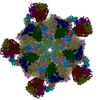 8k42C 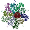 8k43C 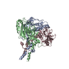 8k44C 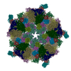 8k49C 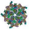 8k4aC 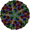 8w9pC 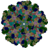 8w9qC C: citing same article ( M: map data used to model this data |
|---|---|
| Similar structure data |


-Links
 PDBj
PDBj- Assembly
| Deposited unit | 
|
|---|---|
| 1 | x 60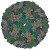
|
| 2 |
|
| 3 | x 5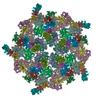
|
| 4 | x 6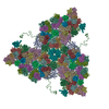
|
| 5 | 
|
| Symmetry | Point symmetry: (Schoenflies symbol: I (icosahedral)) |
-Components
| #1: Protein | Mass: 108031.008 Da / Num. of mol.: 2 / Source method: isolated from a natural source / Details: OR004519 / Source: (natural) Banna virus / References: UniProt: Q9INH3#2: Protein | Mass: 32885.305 Da / Num. of mol.: 13 / Source method: isolated from a natural source / Details: OR004525 / Source: (natural) Banna virus / References: UniProt: W0G587#3: Protein | Mass: 28655.770 Da / Num. of mol.: 2 / Source method: isolated from a natural source / Details: OR004527 / Source: (natural) Banna virus / References: UniProt: A0A2H4QDD3Has protein modification | N | |
|---|
-Experimental details
-Experiment
| Experiment | Method: ELECTRON MICROSCOPY |
|---|---|
| EM experiment | Aggregation state: PARTICLE / 3D reconstruction method: single particle reconstruction |
- Sample preparation
| Component | Name: Banna virus / Type: VIRUS / Entity ID: all / Source: NATURAL |
|---|---|
| Source (natural) | Organism: Banna virus / Strain: YN15-126-01 |
| Details of virus | Empty: NO / Enveloped: NO / Isolate: SPECIES / Type: VIRION |
| Buffer solution | pH: 7.4 |
| Specimen | Embedding applied: NO / Shadowing applied: NO / Staining applied: NO / Vitrification applied: YES |
| Vitrification | Instrument: FEI VITROBOT MARK IV / Cryogen name: ETHANE / Humidity: 100 % / Chamber temperature: 289 K |
- Electron microscopy imaging
| Microscopy | Model: JEOL CRYO ARM 300 |
|---|---|
| Electron gun | Electron source:  FIELD EMISSION GUN / Accelerating voltage: 300 kV / Illumination mode: FLOOD BEAM FIELD EMISSION GUN / Accelerating voltage: 300 kV / Illumination mode: FLOOD BEAM |
| Electron lens | Mode: BRIGHT FIELD / Nominal defocus max: 2500 nm / Nominal defocus min: 500 nm |
| Image recording | Electron dose: 40 e/Å2 / Film or detector model: GATAN K3 (6k x 4k) |
- Processing
| EM software |
| ||||||||||||
|---|---|---|---|---|---|---|---|---|---|---|---|---|---|
| CTF correction | Type: PHASE FLIPPING AND AMPLITUDE CORRECTION | ||||||||||||
| 3D reconstruction | Resolution: 4.8 Å / Resolution method: FSC 0.143 CUT-OFF / Num. of particles: 42352 / Symmetry type: POINT |
