 Movie
Movie Controller


+ Open data

- Basic information
| Entry | Database: PDB / ID: 8w7h | ||||||
|---|---|---|---|---|---|---|---|
| Title | Purine Nucleoside Phosphorylase in complex with MMV000848 | ||||||
 Components Components | Purine nucleoside phosphorylase | ||||||
 Keywords Keywords | TRANSFERASE/INHIBITOR / Protein binding / TRANSFERASE-inhibitor complex | ||||||
| Function / homology |  Function and homology information Function and homology informationPyrimidine salvage / Pyrimidine catabolism / S-methyl-5'-thioinosine phosphorylase / purine nucleotide catabolic process / uridine catabolic process / inosine catabolic process / S-methyl-5-thioadenosine phosphorylase activity / guanosine phosphorylase activity / uridine phosphorylase activity / purine-nucleoside phosphorylase ...Pyrimidine salvage / Pyrimidine catabolism / S-methyl-5'-thioinosine phosphorylase / purine nucleotide catabolic process / uridine catabolic process / inosine catabolic process / S-methyl-5-thioadenosine phosphorylase activity / guanosine phosphorylase activity / uridine phosphorylase activity / purine-nucleoside phosphorylase / purine-nucleoside phosphorylase activity / purine ribonucleoside salvage / cytosol Similarity search - Function | ||||||
| Biological species |  | ||||||
| Method |  X-RAY DIFFRACTION / SYNCHROTRON / MOLECULAR REPLACEMENT / Resolution: 1.85 Å X-RAY DIFFRACTION / SYNCHROTRON / MOLECULAR REPLACEMENT / Resolution: 1.85 Å | ||||||
 Authors Authors | Chung, Z. / Lin, J.Q. / Lescar, J. | ||||||
| Funding support | 1items
| ||||||
 Citation Citation | Journal: J.Biol.Chem. / Year: 2023 Title: Identification and structural validation of purine nucleoside phosphorylase from Plasmodium falciparum as a target of MMV000848. Authors: Chung, Z. / Lin, J. / Wirjanata, G. / Dziekan, J.M. / El Sahili, A. / Preiser, P.R. / Bozdech, Z. / Lescar, J. | ||||||
| History |
|
- Structure visualization
| Structure viewer | Molecule: MolmilJmol/JSmol |
|---|
- Downloads & links
-Download
| PDBx/mmCIF format | 8w7h.cif.gz | 121 KB | Display | PDBx/mmCIF format |
|---|---|---|---|---|
| PDB format | pdb8w7h.ent.gz | 87.8 KB | Display | PDB format |
| PDBx/mmJSON format | 8w7h.json.gz | Tree view | PDBx/mmJSON format | |
| Others |  Other downloads Other downloads |
-Validation report
| Arichive directory | https://data.pdbj.org/pub/pdb/validation_reports/w7/8w7hftp://data.pdbj.org/pub/pdb/validation_reports/w7/8w7h | HTTPS FTP |
|---|
-Related structure data
| Similar structure data |
|---|
-Links
 PDBj
PDBj- Assembly
| Deposited unit | 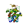
| ||||||||||||||||||||||||
|---|---|---|---|---|---|---|---|---|---|---|---|---|---|---|---|---|---|---|---|---|---|---|---|---|---|
| 1 | x 6
| ||||||||||||||||||||||||
| 2 |
| ||||||||||||||||||||||||
| 3 | 
| ||||||||||||||||||||||||
| 4 | 
| ||||||||||||||||||||||||
| Unit cell |
| ||||||||||||||||||||||||
| Symmetry | Point symmetry: (Schoenflies symbol: D3 (2x3 fold dihedral)) | ||||||||||||||||||||||||
| Components on special symmetry positions |
|
-Components
-Protein , 1 types, 1 molecules A
| #1: Protein | Mass: 27791.117 Da / Num. of mol.: 1 Source method: isolated from a genetically manipulated source Source: (gene. exp.)  |
|---|
-Non-polymers , 5 types, 175 molecules 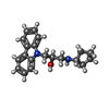


| #2: Chemical | ChemComp-XUH / ( Mass: 308.417 Da / Num. of mol.: 1 / Source method: obtained synthetically / Formula: C20H24N2O / Feature type: SUBJECT OF INVESTIGATION Mass: 308.417 Da / Num. of mol.: 1 / Source method: obtained synthetically / Formula: C20H24N2O / Feature type: SUBJECT OF INVESTIGATION | ||||
|---|---|---|---|---|---|
| #3: Chemical | ChemComp-EDO /  Mass: 62.068 Da / Num. of mol.: 1 / Source method: obtained synthetically / Formula: C2H6O2 Mass: 62.068 Da / Num. of mol.: 1 / Source method: obtained synthetically / Formula: C2H6O2 | ||||
| #4: Chemical |  Mass: 22.990 Da / Num. of mol.: 2 / Source method: obtained synthetically / Formula: Na Mass: 22.990 Da / Num. of mol.: 2 / Source method: obtained synthetically / Formula: Na#5: Chemical | ChemComp-PO4 / |  Mass: 94.971 Da / Num. of mol.: 1 / Source method: obtained synthetically / Formula: PO4 Mass: 94.971 Da / Num. of mol.: 1 / Source method: obtained synthetically / Formula: PO4#6: Water | ChemComp-HOH / | Mass: 18.015 Da / Num. of mol.: 170 / Source method: isolated from a natural source / Formula: H2O |
-Details
| Has ligand of interest | Y |
|---|
-Experimental details
-Experiment
| Experiment | Method: X-RAY DIFFRACTION / Number of used crystals: 1 |
|---|
- Sample preparation
| Crystal | Density Matthews: 2.14 Å3/Da / Density % sol: 42.61 % |
|---|---|
| Crystal grow | Temperature: 293.15 K / Method: vapor diffusion, sitting drop / pH: 7.5 / Details: 10% (w/v) PEG 1000, 10% (w/v) PEG 8000 |
-Data collection
| Diffraction | Mean temperature: 80 K / Ambient temp details: Nitrogen gas / Serial crystal experiment: N |
|---|---|
| Diffraction source | Source: SYNCHROTRON / Site: Australian Synchrotron  / Beamline: MX1 / Wavelength: 0.9537 Å / Beamline: MX1 / Wavelength: 0.9537 Å |
| Detector | Type: DECTRIS EIGER2 X 9M / Detector: PIXEL / Date: Apr 23, 2023 |
| Radiation | Protocol: SINGLE WAVELENGTH / Monochromatic (M) / Laue (L): M / Scattering type: x-ray |
| Radiation wavelength | Wavelength: 0.9537 Å / Relative weight: 1 |
| Reflection | Resolution: 1.85→33.17 Å / Num. obs: 20977 / % possible obs: 99.79 % / Redundancy: 20.3 % / Biso Wilson estimate: 27.17 Å2 / CC1/2: 1 / CC star: 1 / Rmerge(I) obs: 0.0743 / Rpim(I) all: 0.01689 / Rrim(I) all: 0.07623 / Net I/σ(I): 30.42 |
| Reflection shell | Resolution: 1.85→1.916 Å / Redundancy: 20.7 % / Rmerge(I) obs: 0.9243 / Mean I/σ(I) obs: 3.73 / Num. unique obs: 2065 / CC1/2: 0.953 / CC star: 0.988 / Rpim(I) all: 0.2069 / Rrim(I) all: 0.9475 / % possible all: 99.32 |
- Processing
| Software |
| |||||||||||||||||||||||||||||||||||||||||||||||||||||||||||||||||||||||||||||||||||||||||||||||||||||||||
|---|---|---|---|---|---|---|---|---|---|---|---|---|---|---|---|---|---|---|---|---|---|---|---|---|---|---|---|---|---|---|---|---|---|---|---|---|---|---|---|---|---|---|---|---|---|---|---|---|---|---|---|---|---|---|---|---|---|---|---|---|---|---|---|---|---|---|---|---|---|---|---|---|---|---|---|---|---|---|---|---|---|---|---|---|---|---|---|---|---|---|---|---|---|---|---|---|---|---|---|---|---|---|---|---|---|---|
| Refinement | Method to determine structure: MOLECULAR REPLACEMENT / Resolution: 1.85→33.17 Å / SU ML: 0.2082 / Cross valid method: FREE R-VALUE / σ(F): 1.33 / Phase error: 25.3595 Stereochemistry target values: GeoStd + Monomer Library + CDL v1.2
| |||||||||||||||||||||||||||||||||||||||||||||||||||||||||||||||||||||||||||||||||||||||||||||||||||||||||
| Solvent computation | Shrinkage radii: 0.9 Å / VDW probe radii: 1.11 Å / Solvent model: FLAT BULK SOLVENT MODEL | |||||||||||||||||||||||||||||||||||||||||||||||||||||||||||||||||||||||||||||||||||||||||||||||||||||||||
| Displacement parameters | Biso mean: 39.11 Å2 | |||||||||||||||||||||||||||||||||||||||||||||||||||||||||||||||||||||||||||||||||||||||||||||||||||||||||
| Refinement step | Cycle: LAST / Resolution: 1.85→33.17 Å
| |||||||||||||||||||||||||||||||||||||||||||||||||||||||||||||||||||||||||||||||||||||||||||||||||||||||||
| Refine LS restraints |
| |||||||||||||||||||||||||||||||||||||||||||||||||||||||||||||||||||||||||||||||||||||||||||||||||||||||||
| LS refinement shell |
|
