 Movie
Movie Controller

[English] 日本語
 Yorodumi
- PDB-8w4n: Crystal structure of EndoSz mutant D234M, in space group P21, in ... -
+ Open data

- Basic information
| Entry | Database: PDB / ID: 8w4n | ||||||
|---|---|---|---|---|---|---|---|
| Title | Crystal structure of EndoSz mutant D234M, in space group P21, in complex with oligosaccharide G2S1 | ||||||
 Components Components | Glycoside hydrolase | ||||||
 Keywords Keywords | HYDROLASE / N-linked glycan / glycoside hydrolase / transglycosylation | ||||||
| Biological species |  Streptococcus equi subsp. zooepidemicus Sz105 (bacteria) Streptococcus equi subsp. zooepidemicus Sz105 (bacteria) | ||||||
| Method |  X-RAY DIFFRACTION / SYNCHROTRON / MOLECULAR REPLACEMENT / Resolution: 3.1 Å X-RAY DIFFRACTION / SYNCHROTRON / MOLECULAR REPLACEMENT / Resolution: 3.1 Å | ||||||
 Authors Authors | Guan, H.H. / Lin, C.C. / Hsieh, Y.C. / Chen, C.J. | ||||||
| Funding support |  Taiwan, 1items Taiwan, 1items
| ||||||
 Citation Citation | Journal: Jacs Au / Year: 2024 Title: Structure-Based High-Efficiency Homogeneous Antibody Platform by Endoglycosidase Sz Provides Insights into Its Transglycosylation Mechanism. Authors: Hsieh, Y.C. / Guan, H.H. / Lin, C.C. / Huang, T.Y. / Chuankhayan, P. / Chen, N.C. / Wang, N.H. / Hu, P.L. / Tsai, Y.C. / Huang, Y.C. / Yoshimura, M. / Lin, P.J. / Hsieh, Y.H. / Chen, C.J. | ||||||
| History |
|
- Structure visualization
| Structure viewer | Molecule:  MolmilJmol/JSmol MolmilJmol/JSmol |
|---|
- Downloads & links
-Download
| PDBx/mmCIF format | 8w4n.cif.gz | 194.3 KB | Display | PDBx/mmCIF format |
|---|---|---|---|---|
| PDB format | pdb8w4n.ent.gz | 147.1 KB | Display | PDB format |
| PDBx/mmJSON format | 8w4n.json.gz | Tree view | PDBx/mmJSON format | |
| Others |  Other downloads Other downloads |
-Validation report
| Arichive directory | https://data.pdbj.org/pub/pdb/validation_reports/w4/8w4nftp://data.pdbj.org/pub/pdb/validation_reports/w4/8w4n | HTTPS FTP |
|---|
-Related structure data
| Related structure data |  8w4gC 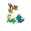 8w4iC 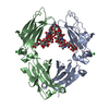 8w4lC 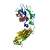 8w4mC  8x8gC  8w4k C: citing same article ( |
|---|
-Links
 PDBj
PDBj- Assembly
| Deposited unit | 
| ||||||||
|---|---|---|---|---|---|---|---|---|---|
| 1 |
| ||||||||
| Unit cell |
|
-Components
| #1: Protein | Mass: 110575.906 Da / Num. of mol.: 1 / Mutation: D234M Source method: isolated from a genetically manipulated source Source: (gene. exp.) Streptococcus equi subsp. zooepidemicus Sz105 (bacteria)Production host: |
|---|---|
| #2: Polysaccharide | N-acetyl-alpha-neuraminic acid-(2-6)-beta-D-galactopyranose-(1-4)-2-acetamido-2-deoxy-beta-D- ...N-acetyl-alpha-neuraminic acid-(2-6)-beta-D-galactopyranose-(1-4)-2-acetamido-2-deoxy-beta-D-glucopyranose-(1-2)-alpha-D-mannopyranose-(1-3)-[beta-D-galactopyranose-(1-4)-2-acetamido-2-deoxy-beta-D-glucopyranose-(1-2)-alpha-D-mannopyranose-(1-6)]beta-D-mannopyranose-(1-4)-2-acetamido-2-deoxy-beta-D-glucopyranose Type: oligosaccharide / Mass: 1729.552 Da / Num. of mol.: 1 Source method: isolated from a genetically manipulated source |
| #3: Chemical | ChemComp-CA /   Mass: 40.078 Da / Num. of mol.: 1 / Source method: obtained synthetically / Formula: Ca Mass: 40.078 Da / Num. of mol.: 1 / Source method: obtained synthetically / Formula: Ca |
| Has ligand of interest | N |
-Experimental details
-Experiment
| Experiment | Method: X-RAY DIFFRACTION / Number of used crystals: 1 |
|---|
- Sample preparation
| Crystal | Density Matthews: 3.08 Å3/Da / Density % sol: 60.05 % |
|---|---|
| Crystal grow | Temperature: 291 K / Method: vapor diffusion, hanging drop Details: 0.1 mM sodium citrate pH 5.4 and 2.2 M ammonium sulfate |
-Data collection
| Diffraction | Mean temperature: 100 K / Serial crystal experiment: N |
|---|---|
| Diffraction source | Source: SYNCHROTRON / Site: NSRRC / Beamline: TPS 05A / Wavelength: 1 Å |
| Detector | Type: RAYONIX MX300-HS / Detector: CCD / Date: Aug 14, 2019 |
| Radiation | Protocol: SINGLE WAVELENGTH / Monochromatic (M) / Laue (L): M / Scattering type: x-ray |
| Radiation wavelength | Wavelength: 1 Å / Relative weight: 1 |
| Reflection | Resolution: 3.1→28.33 Å / Num. obs: 24061 / % possible obs: 98.23 % / Redundancy: 1.8 % / CC1/2: 0.988 / Net I/σ(I): 5 |
| Reflection shell | Resolution: 3.1→3.21 Å / Num. unique obs: 2397 / CC1/2: 0.54 |
- Processing
| Software |
| |||||||||||||||||||||||||||||||||||||||||||||||||||||||||||||||||||||||||||||||||||||||||||||||||||||||||
|---|---|---|---|---|---|---|---|---|---|---|---|---|---|---|---|---|---|---|---|---|---|---|---|---|---|---|---|---|---|---|---|---|---|---|---|---|---|---|---|---|---|---|---|---|---|---|---|---|---|---|---|---|---|---|---|---|---|---|---|---|---|---|---|---|---|---|---|---|---|---|---|---|---|---|---|---|---|---|---|---|---|---|---|---|---|---|---|---|---|---|---|---|---|---|---|---|---|---|---|---|---|---|---|---|---|---|
| Refinement | Method to determine structure: MOLECULAR REPLACEMENT / Resolution: 3.1→28.33 Å / SU ML: 0.53 / Cross valid method: THROUGHOUT / σ(F): 1.35 / Phase error: 31.2 / Stereochemistry target values: ML
| |||||||||||||||||||||||||||||||||||||||||||||||||||||||||||||||||||||||||||||||||||||||||||||||||||||||||
| Solvent computation | Shrinkage radii: 0.9 Å / VDW probe radii: 1.11 Å / Solvent model: FLAT BULK SOLVENT MODEL | |||||||||||||||||||||||||||||||||||||||||||||||||||||||||||||||||||||||||||||||||||||||||||||||||||||||||
| Refinement step | Cycle: LAST / Resolution: 3.1→28.33 Å
| |||||||||||||||||||||||||||||||||||||||||||||||||||||||||||||||||||||||||||||||||||||||||||||||||||||||||
| Refine LS restraints |
| |||||||||||||||||||||||||||||||||||||||||||||||||||||||||||||||||||||||||||||||||||||||||||||||||||||||||
| LS refinement shell |
|
