 Movie
Movie Controller

[English] 日本語
 Yorodumi
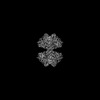
- PDB-8w2j: Human liver phosphofructokinase-1 filament in the T-state conformation -
+ Open data

- Basic information
| Entry | Database: PDB / ID: 8w2j | ||||||
|---|---|---|---|---|---|---|---|
| Title | Human liver phosphofructokinase-1 filament in the T-state conformation | ||||||
 Components Components | ATP-dependent 6-phosphofructokinase, liver type | ||||||
 Keywords Keywords | TRANSFERASE / PFK / glycolysis | ||||||
| Function / homology |  Function and homology information Function and homology information6-phosphofructokinase complex / 6-phosphofructokinase / 6-phosphofructokinase activity / fructose binding / fructose-6-phosphate binding / fructose 1,6-bisphosphate metabolic process / fructose 6-phosphate metabolic process / Glycolysis / canonical glycolysis / negative regulation of insulin secretion ...6-phosphofructokinase complex / 6-phosphofructokinase / 6-phosphofructokinase activity / fructose binding / fructose-6-phosphate binding / fructose 1,6-bisphosphate metabolic process / fructose 6-phosphate metabolic process / Glycolysis / canonical glycolysis / negative regulation of insulin secretion / response to glucose / glycolytic process / kinase binding / secretory granule lumen / ficolin-1-rich granule lumen / Neutrophil degranulation / extracellular exosome / extracellular region / ATP binding / membrane / metal ion binding / identical protein binding / cytosol Similarity search - Function | ||||||
| Biological species |  Homo sapiens (human) Homo sapiens (human) | ||||||
| Method | ELECTRON MICROSCOPY / single particle reconstruction / cryo EM / Resolution: 3.1 Å | ||||||
 Authors Authors | Lynch, E.M. / Kollman, J.M. / Webb, B.A. | ||||||
| Funding support |  United States, 1items United States, 1items
| ||||||
 Citation Citation | Journal: Nat Commun / Year: 2024 Title: Structural basis for allosteric regulation of human phosphofructokinase-1. Authors: Eric M Lynch / Heather Hansen / Lauren Salay / Madison Cooper / Stepan Timr / Justin M Kollman / Bradley A Webb /  Abstract: Phosphofructokinase-1 (PFK1) catalyzes the rate-limiting step of glycolysis, committing glucose to conversion into cellular energy. PFK1 is highly regulated to respond to the changing energy needs of ...Phosphofructokinase-1 (PFK1) catalyzes the rate-limiting step of glycolysis, committing glucose to conversion into cellular energy. PFK1 is highly regulated to respond to the changing energy needs of the cell. In bacteria, the structural basis of PFK1 regulation is a textbook example of allostery; molecular signals of low and high cellular energy promote transition between an active R-state and inactive T-state conformation, respectively. Little is known, however, about the structural basis for regulation of eukaryotic PFK1. Here, we determine structures of the human liver isoform of PFK1 (PFKL) in the R- and T-state by cryoEM, providing insight into eukaryotic PFK1 allosteric regulatory mechanisms. The T-state structure reveals conformational differences between the bacterial and eukaryotic enzyme, the mechanisms of allosteric inhibition by ATP binding at multiple sites, and an autoinhibitory role of the C-terminus in stabilizing the T-state. We also determine structures of PFKL filaments that define the mechanism of higher-order assembly and demonstrate that these structures are necessary for higher-order assembly of PFKL in cells. | ||||||
| History |
|
- Structure visualization
| Structure viewer | Molecule: MolmilJmol/JSmol |
|---|
- Downloads & links
-Download
| PDBx/mmCIF format | 8w2j.cif.gz | 1.8 MB | Display | PDBx/mmCIF format |
|---|---|---|---|---|
| PDB format | pdb8w2j.ent.gz | 1.6 MB | Display | PDB format |
| PDBx/mmJSON format | 8w2j.json.gz | Tree view | PDBx/mmJSON format | |
| Others |  Other downloads Other downloads |
-Validation report
| Arichive directory | https://data.pdbj.org/pub/pdb/validation_reports/w2/8w2jftp://data.pdbj.org/pub/pdb/validation_reports/w2/8w2j | HTTPS FTP |
|---|
-Related structure data
| Related structure data | 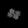 43750MC 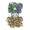 8w2gC 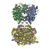 8w2hC  8w2iC M: map data used to model this data C: citing same article ( |
|---|---|
| Similar structure data |


-Links
 PDBj
PDBj


- Assembly
| Deposited unit | 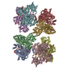
|
|---|---|
| 1 |
|
-Components
| #1: Protein | Mass: 85120.375 Da / Num. of mol.: 8 Source method: isolated from a genetically manipulated source Source: (gene. exp.) Homo sapiens (human) / Gene: PFKL / Production host:  unidentified baculovirus / References: UniProt: P17858, 6-phosphofructokinase unidentified baculovirus / References: UniProt: P17858, 6-phosphofructokinase#2: Sugar | ChemComp-FBP / 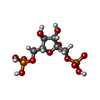  Type: D-saccharide, beta linking / Mass: 340.116 Da / Num. of mol.: 8 / Source method: obtained synthetically / Formula: C6H14O12P2 / Feature type: SUBJECT OF INVESTIGATION Type: D-saccharide, beta linking / Mass: 340.116 Da / Num. of mol.: 8 / Source method: obtained synthetically / Formula: C6H14O12P2 / Feature type: SUBJECT OF INVESTIGATION#3: Chemical | ChemComp-ATP /   Mass: 507.181 Da / Num. of mol.: 24 / Source method: obtained synthetically / Formula: C10H16N5O13P3 / Feature type: SUBJECT OF INVESTIGATION / Comment: ATP, energy-carrying molecule*YM Mass: 507.181 Da / Num. of mol.: 24 / Source method: obtained synthetically / Formula: C10H16N5O13P3 / Feature type: SUBJECT OF INVESTIGATION / Comment: ATP, energy-carrying molecule*YM#4: Chemical | ChemComp-MG /   Mass: 24.305 Da / Num. of mol.: 8 / Source method: obtained synthetically / Formula: Mg Mass: 24.305 Da / Num. of mol.: 8 / Source method: obtained synthetically / Formula: MgHas ligand of interest | Y | |
|---|
-Experimental details
-Experiment
| Experiment | Method: ELECTRON MICROSCOPY |
|---|---|
| EM experiment | Aggregation state: PARTICLE / 3D reconstruction method: single particle reconstruction |
- Sample preparation
| Component | Name: PFKL filament / Type: COMPLEX / Entity ID: #1 / Source: RECOMBINANT |
|---|---|
| Molecular weight | Experimental value: NO |
| Source (natural) | Organism: Homo sapiens (human) |
| Source (recombinant) | Organism: unidentified baculovirus |
| Buffer solution | pH: 7.5 |
| Specimen | Embedding applied: NO / Shadowing applied: NO / Staining applied: NO / Vitrification applied: YES |
| Vitrification | Cryogen name: ETHANE / Humidity: 100 % |
- Electron microscopy imaging
| Experimental equipment |  Model: Titan Krios / Image courtesy: FEI Company |
|---|---|
| Microscopy | Model: FEI TITAN KRIOS |
| Electron gun | Electron source:  FIELD EMISSION GUN / Accelerating voltage: 300 kV / Illumination mode: FLOOD BEAM FIELD EMISSION GUN / Accelerating voltage: 300 kV / Illumination mode: FLOOD BEAM |
| Electron lens | Mode: BRIGHT FIELD / Nominal magnification: 130000 X / Nominal defocus max: 2400 nm / Nominal defocus min: 800 nm |
| Specimen holder | Cryogen: NITROGEN |
| Image recording | Electron dose: 90 e/Å2 / Film or detector model: GATAN K2 SUMMIT (4k x 4k) |
- Processing
| EM software |
| |||||||||||||||||||||||||||||||||
|---|---|---|---|---|---|---|---|---|---|---|---|---|---|---|---|---|---|---|---|---|---|---|---|---|---|---|---|---|---|---|---|---|---|---|
| CTF correction | Type: PHASE FLIPPING AND AMPLITUDE CORRECTION | |||||||||||||||||||||||||||||||||
| 3D reconstruction | Resolution: 3.1 Å / Resolution method: FSC 0.143 CUT-OFF / Num. of particles: 45362 / Symmetry type: POINT | |||||||||||||||||||||||||||||||||
| Atomic model building | Protocol: FLEXIBLE FIT / Space: REAL | |||||||||||||||||||||||||||||||||
| Atomic model building | Source name: Modeller / Type: in silico model |
