 Movie
Movie Controller

[English] 日本語
 Yorodumi
- PDB-8w1q: Aerobic crystal structure of iron-bound FlcD from Pseudomonas aer... -
+ Open data

- Basic information
| Entry | Database: PDB / ID: 8w1q | |||||||||
|---|---|---|---|---|---|---|---|---|---|---|
| Title | Aerobic crystal structure of iron-bound FlcD from Pseudomonas aeruginosa | |||||||||
 Components Components | Pyrroloquinoline quinone (Coenzyme PQQ) biosynthesis protein C | |||||||||
 Keywords Keywords | OXIDOREDUCTASE / mono-iron dioxygenase | |||||||||
| Function / homology |  Function and homology information Function and homology informationpyrroloquinoline-quinone synthase / hydrolase activity, hydrolyzing O-glycosyl compounds / periplasmic space / oxidoreductase activity / metal ion binding Similarity search - Function | |||||||||
| Biological species |   Pseudomonas aeruginosa (bacteria) Pseudomonas aeruginosa (bacteria) | |||||||||
| Method |  X-RAY DIFFRACTION / SYNCHROTRON / MOLECULAR REPLACEMENT / Resolution: 1.56 Å X-RAY DIFFRACTION / SYNCHROTRON / MOLECULAR REPLACEMENT / Resolution: 1.56 Å | |||||||||
 Authors Authors | Walker, M.E. / Grove, T.L. / Li, B. / Redinbo, M.R. | |||||||||
| Funding support |  United States, 2items United States, 2items
| |||||||||
 Citation Citation | Journal: Acs Cent.Sci. / Year: 2024 Title: Structural Basis for Methine Excision by a Heme Oxygenase-like Enzyme. Authors: Simke, W.C. / Walker, M.E. / Calderone, L.A. / Putz, A.T. / Patteson, J.B. / Vitro, C.N. / Zizola, C.F. / Redinbo, M.R. / Pandelia, M.E. / Grove, T.L. / Li, B. | |||||||||
| History |
|
- Structure visualization
| Structure viewer | Molecule: MolmilJmol/JSmol |
|---|
- Downloads & links
-Download
| PDBx/mmCIF format | 8w1q.cif.gz | 189 KB | Display | PDBx/mmCIF format |
|---|---|---|---|---|
| PDB format | pdb8w1q.ent.gz | 118.7 KB | Display | PDB format |
| PDBx/mmJSON format | 8w1q.json.gz | Tree view | PDBx/mmJSON format | |
| Others |  Other downloads Other downloads |
-Validation report
| Summary document | 8w1q_validation.pdf.gz | 1.5 MB | Display | wwPDB validaton report |
|---|---|---|---|---|
| Full document | 8w1q_full_validation.pdf.gz | 1.5 MB | Display | |
| Data in XML | 8w1q_validation.xml.gz | 29.2 KB | Display | |
| Data in CIF | 8w1q_validation.cif.gz | 44.1 KB | Display | |
| Arichive directory | https://data.pdbj.org/pub/pdb/validation_reports/w1/8w1qftp://data.pdbj.org/pub/pdb/validation_reports/w1/8w1q | HTTPS FTP |
-Related structure data


-Links
 PDBj
PDBj


- Assembly
| Deposited unit | 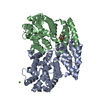
| ||||||||||||
|---|---|---|---|---|---|---|---|---|---|---|---|---|---|
| 1 |
| ||||||||||||
| Unit cell |
|
-Components
-Protein , 1 types, 2 molecules AB
| #1: Protein | Mass: 40240.246 Da / Num. of mol.: 2 Source method: isolated from a genetically manipulated source Source: (gene. exp.) Pseudomonas aeruginosa (bacteria)Gene: pqqC_2, CAZ10_19980, DT376_18990, NCTC13621_03444, PAERUG_P19_London_7_VIM_2_05_10_05324 Production host: References: UniProt: A0A0C7AN42, pyrroloquinoline-quinone synthase |
|---|
-Non-polymers , 5 types, 552 molecules 


| #2: Chemical |  Mass: 55.845 Da / Num. of mol.: 2 / Source method: obtained synthetically / Formula: Fe / Feature type: SUBJECT OF INVESTIGATION Mass: 55.845 Da / Num. of mol.: 2 / Source method: obtained synthetically / Formula: Fe / Feature type: SUBJECT OF INVESTIGATION#3: Chemical |  Mass: 39.098 Da / Num. of mol.: 2 / Source method: obtained synthetically / Formula: K Mass: 39.098 Da / Num. of mol.: 2 / Source method: obtained synthetically / Formula: K#4: Chemical | ChemComp-MG / |  Mass: 24.305 Da / Num. of mol.: 1 / Source method: obtained synthetically / Formula: Mg Mass: 24.305 Da / Num. of mol.: 1 / Source method: obtained synthetically / Formula: Mg#5: Chemical | ChemComp-GOL / |  Mass: 92.094 Da / Num. of mol.: 1 / Source method: obtained synthetically / Formula: C3H8O3 Mass: 92.094 Da / Num. of mol.: 1 / Source method: obtained synthetically / Formula: C3H8O3#6: Water | ChemComp-HOH / | Mass: 18.015 Da / Num. of mol.: 546 / Source method: isolated from a natural source / Formula: H2O |
|---|
-Details
| Has ligand of interest | Y |
|---|
-Experimental details
-Experiment
| Experiment | Method: X-RAY DIFFRACTION / Number of used crystals: 1 |
|---|
- Sample preparation
| Crystal | Density % sol: 33.78 % |
|---|---|
| Crystal grow | Temperature: 293 K / Method: vapor diffusion, sitting drop / Details: 0.2 M magnesium chloride, 20% w/v PEG 3350 |
-Data collection
| Diffraction | Mean temperature: 100 K / Serial crystal experiment: N |
|---|---|
| Diffraction source | Source: SYNCHROTRON / Site: APS / Beamline: 23-ID-D / Wavelength: 1 Å |
| Detector | Type: DECTRIS PILATUS3 6M / Detector: PIXEL / Date: Nov 14, 2020 |
| Radiation | Protocol: SINGLE WAVELENGTH / Monochromatic (M) / Laue (L): M / Scattering type: x-ray |
| Radiation wavelength | Wavelength: 1 Å / Relative weight: 1 |
| Reflection | Resolution: 1.56→31.57 Å / Num. obs: 79607 / % possible obs: 95.19 % / Redundancy: 3 % / Biso Wilson estimate: 18.17 Å2 / CC1/2: 0.992 / CC star: 0.998 / Rmerge(I) obs: 0.09696 / Rpim(I) all: 0.0637 / Rrim(I) all: 0.1166 / Net I/σ(I): 6.46 |
| Reflection shell | Resolution: 1.56→1.616 Å / Redundancy: 2.4 % / Rmerge(I) obs: 0.4948 / Mean I/σ(I) obs: 1.32 / Num. unique obs: 7453 / CC1/2: 0.508 / CC star: 0.821 / Rpim(I) all: 0.373 / Rrim(I) all: 0.6235 / % possible all: 89.23 |
- Processing
| Software |
| |||||||||||||||||||||||||||||||||||||||||||||||||||||||||||||||||||||||||||||||||||||||||||||||||||||||||
|---|---|---|---|---|---|---|---|---|---|---|---|---|---|---|---|---|---|---|---|---|---|---|---|---|---|---|---|---|---|---|---|---|---|---|---|---|---|---|---|---|---|---|---|---|---|---|---|---|---|---|---|---|---|---|---|---|---|---|---|---|---|---|---|---|---|---|---|---|---|---|---|---|---|---|---|---|---|---|---|---|---|---|---|---|---|---|---|---|---|---|---|---|---|---|---|---|---|---|---|---|---|---|---|---|---|---|
| Refinement | Method to determine structure: MOLECULAR REPLACEMENT / Resolution: 1.56→31.57 Å / SU ML: 0.1861 / Cross valid method: FREE R-VALUE / σ(F): 1.34 / Phase error: 22.4771 Stereochemistry target values: GeoStd + Monomer Library + CDL v1.2
| |||||||||||||||||||||||||||||||||||||||||||||||||||||||||||||||||||||||||||||||||||||||||||||||||||||||||
| Solvent computation | Shrinkage radii: 0.9 Å / VDW probe radii: 1.11 Å / Solvent model: FLAT BULK SOLVENT MODEL | |||||||||||||||||||||||||||||||||||||||||||||||||||||||||||||||||||||||||||||||||||||||||||||||||||||||||
| Displacement parameters | Biso mean: 25.44 Å2 | |||||||||||||||||||||||||||||||||||||||||||||||||||||||||||||||||||||||||||||||||||||||||||||||||||||||||
| Refinement step | Cycle: LAST / Resolution: 1.56→31.57 Å
| |||||||||||||||||||||||||||||||||||||||||||||||||||||||||||||||||||||||||||||||||||||||||||||||||||||||||
| Refine LS restraints |
| |||||||||||||||||||||||||||||||||||||||||||||||||||||||||||||||||||||||||||||||||||||||||||||||||||||||||
| LS refinement shell |
|
