 Movie
Movie Controller

[English] 日本語
 Yorodumi
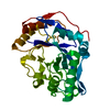
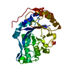
- PDB-8w04: Crystal structure of DUF1735-domain containing protein (GH18-like... -
+ Open data

- Basic information
| Entry | Database: PDB / ID: 8w04 | ||||||
|---|---|---|---|---|---|---|---|
| Title | Crystal structure of DUF1735-domain containing protein (GH18-like) from Bacteroides faecium at 2.9 A resolution (Space group P21) | ||||||
 Components Components | DUF1735 domain-containing protein | ||||||
 Keywords Keywords | SUGAR BINDING PROTEIN / DUF1735-domain containing protein / GH18-like / inactive endoglycosidase / Bacteroides faecium / gut microbiome | ||||||
| Function / homology | Domain of unknown function DUF1735 / BT_3987-like, N-terminal domain / Glycosyl hydrolases family 18 / Glycoside hydrolase family 18, catalytic domain / Prokaryotic membrane lipoprotein lipid attachment site profile. / Glycoside hydrolase superfamily / carbohydrate metabolic process / DUF1735 domain-containing protein Function and homology information Function and homology information | ||||||
| Biological species |  Bacteroides faecium (bacteria) Bacteroides faecium (bacteria) | ||||||
| Method |  X-RAY DIFFRACTION / SYNCHROTRON / MOLECULAR REPLACEMENT / Resolution: 2.91 Å X-RAY DIFFRACTION / SYNCHROTRON / MOLECULAR REPLACEMENT / Resolution: 2.91 Å | ||||||
 Authors Authors | Sastre, D.E. / Navarro, M.V.A.S. / Sultana, N. / Sundberg, E.J. | ||||||
| Funding support |  United States, 1items United States, 1items
| ||||||
 Citation Citation | Journal: Nat Commun / Year: 2024 Title: Human gut microbes express functionally distinct endoglycosidases to metabolize the same N-glycan substrate. Authors: Sastre, D.E. / Sultana, N. / V A S Navarro, M. / Huliciak, M. / Du, J. / Cifuente, J.O. / Flowers, M. / Liu, X. / Lollar, P. / Trastoy, B. / Guerin, M.E. / Sundberg, E.J. | ||||||
| History |
|
- Structure visualization
| Structure viewer | Molecule: MolmilJmol/JSmol |
|---|
- Downloads & links
-Download
| PDBx/mmCIF format | 8w04.cif.gz | 187.3 KB | Display | PDBx/mmCIF format |
|---|---|---|---|---|
| PDB format | pdb8w04.ent.gz | 146.5 KB | Display | PDB format |
| PDBx/mmJSON format | 8w04.json.gz | Tree view | PDBx/mmJSON format | |
| Others |  Other downloads Other downloads |
-Validation report
| Arichive directory | https://data.pdbj.org/pub/pdb/validation_reports/w0/8w04ftp://data.pdbj.org/pub/pdb/validation_reports/w0/8w04 | HTTPS FTP |
|---|
-Related structure data


-Links
 PDBj
PDBj


- Assembly
| Deposited unit | 
| ||||||||
|---|---|---|---|---|---|---|---|---|---|
| 1 |
| ||||||||
| Unit cell |
|
-Components
| #1: Protein | Mass: 57349.098 Da / Num. of mol.: 2 Source method: isolated from a genetically manipulated source Source: (gene. exp.) Bacteroides faecium (bacteria) / Gene: BacF7301_21000 / Production host: |
|---|
-Experimental details
-Experiment
| Experiment | Method: X-RAY DIFFRACTION / Number of used crystals: 1 |
|---|
- Sample preparation
| Crystal | Density Matthews: 2.55 Å3/Da / Density % sol: 51.83 % |
|---|---|
| Crystal grow | Temperature: 297 K / Method: vapor diffusion, sitting drop Details: 0.2 M Sodium bromide, 0.1 M Bis Tris propane pH 7.5, 20% (w/v) PEG 3350 (PACT 2-26 (G2)) |
-Data collection
| Diffraction | Mean temperature: 100 K / Serial crystal experiment: N |
|---|---|
| Diffraction source | Source: SYNCHROTRON / Site: APS / Beamline: 22-BM / Wavelength: 1 Å |
| Detector | Type: RAYONIX MX225-HS / Detector: CCD / Date: Sep 21, 2022 |
| Radiation | Protocol: SINGLE WAVELENGTH / Monochromatic (M) / Laue (L): M / Scattering type: x-ray |
| Radiation wavelength | Wavelength: 1 Å / Relative weight: 1 |
| Reflection | Resolution: 2.9→50 Å / Num. obs: 25271 / % possible obs: 97.72 % / Redundancy: 7.2 % / Biso Wilson estimate: 49.6 Å2 / CC1/2: 0.972 / CC star: 0.993 / Rmerge(I) obs: 0.192 / Rpim(I) all: 0.076 / Rrim(I) all: 0.207 / Χ2: 2.142 / Net I/σ(I): 13.39 |
| Reflection shell | Resolution: 2.9→2.95 Å / Redundancy: 6.8 % / Rmerge(I) obs: 0.773 / Mean I/σ(I) obs: 2.26 / Num. unique obs: 2050 / CC1/2: 0.814 / CC star: 0.947 / Rpim(I) all: 0.315 / Rrim(I) all: 0.836 / Χ2: 0.781 / % possible all: 79.94 |
- Processing
| Software |
| |||||||||||||||||||||||||||||||||||||||||||||||||||||||||||||||||||||||||||||||||||||||||||||||||||||||||
|---|---|---|---|---|---|---|---|---|---|---|---|---|---|---|---|---|---|---|---|---|---|---|---|---|---|---|---|---|---|---|---|---|---|---|---|---|---|---|---|---|---|---|---|---|---|---|---|---|---|---|---|---|---|---|---|---|---|---|---|---|---|---|---|---|---|---|---|---|---|---|---|---|---|---|---|---|---|---|---|---|---|---|---|---|---|---|---|---|---|---|---|---|---|---|---|---|---|---|---|---|---|---|---|---|---|---|
| Refinement | Method to determine structure: MOLECULAR REPLACEMENT / Resolution: 2.91→37.11 Å / SU ML: 0.39 / Cross valid method: FREE R-VALUE / σ(F): 1.34 / Phase error: 26.55 / Stereochemistry target values: ML
| |||||||||||||||||||||||||||||||||||||||||||||||||||||||||||||||||||||||||||||||||||||||||||||||||||||||||
| Solvent computation | Shrinkage radii: 0.9 Å / VDW probe radii: 1.1 Å / Solvent model: FLAT BULK SOLVENT MODEL | |||||||||||||||||||||||||||||||||||||||||||||||||||||||||||||||||||||||||||||||||||||||||||||||||||||||||
| Refinement step | Cycle: LAST / Resolution: 2.91→37.11 Å
| |||||||||||||||||||||||||||||||||||||||||||||||||||||||||||||||||||||||||||||||||||||||||||||||||||||||||
| Refine LS restraints |
| |||||||||||||||||||||||||||||||||||||||||||||||||||||||||||||||||||||||||||||||||||||||||||||||||||||||||
| LS refinement shell |
|
