 Movie
Movie Controller


+ Open data

- Basic information
| Entry | Database: PDB / ID: 8vyr | ||||||
|---|---|---|---|---|---|---|---|
| Title | Cryo-EM Structure of the BRAF V600E monomer bound to GDC0879 | ||||||
 Components Components |
| ||||||
 Keywords Keywords | TRANSFERASE/INHIBITOR / BRAF Kinase Oncogenic Mutant Monomer / TRANSFERASE-INHIBITOR complex | ||||||
| Function / homology |  Function and homology information Function and homology informationsynaptic target recognition / Golgi reassembly / CD4-positive, alpha-beta T cell differentiation / NOTCH4 Activation and Transmission of Signal to the Nucleus / positive regulation of axon regeneration / CD4-positive or CD8-positive, alpha-beta T cell lineage commitment / negative regulation of synaptic vesicle exocytosis / establishment of Golgi localization / respiratory system process / Signalling to p38 via RIT and RIN ...synaptic target recognition / Golgi reassembly / CD4-positive, alpha-beta T cell differentiation / NOTCH4 Activation and Transmission of Signal to the Nucleus / positive regulation of axon regeneration / CD4-positive or CD8-positive, alpha-beta T cell lineage commitment / negative regulation of synaptic vesicle exocytosis / establishment of Golgi localization / respiratory system process / Signalling to p38 via RIT and RIN / head morphogenesis / endothelial cell apoptotic process / myeloid progenitor cell differentiation / ARMS-mediated activation / tube formation / negative regulation of fibroblast migration / SHOC2 M1731 mutant abolishes MRAS complex function / Gain-of-function MRAS complexes activate RAF signaling / regulation of synapse maturation / Rap1 signalling / positive regulation of D-glucose transmembrane transport / establishment of protein localization to membrane / positive regulation of axonogenesis / regulation of T cell differentiation / negative regulation of protein localization to nucleus / KSRP (KHSRP) binds and destabilizes mRNA / Negative feedback regulation of MAPK pathway / GP1b-IX-V activation signalling / Frs2-mediated activation / face development / stress fiber assembly / thyroid gland development / MAP kinase kinase activity / somatic stem cell population maintenance / Regulation of localization of FOXO transcription factors / Interleukin-3, Interleukin-5 and GM-CSF signaling / synaptic vesicle exocytosis / positive regulation of peptidyl-serine phosphorylation / phosphoserine residue binding / Activation of BAD and translocation to mitochondria / negative regulation of endothelial cell apoptotic process / MAP kinase kinase kinase activity / regulation of ERK1 and ERK2 cascade / ERK1 and ERK2 cascade / SARS-CoV-2 targets host intracellular signalling and regulatory pathways / postsynaptic modulation of chemical synaptic transmission / cellular response to glucose starvation / Chk1/Chk2(Cds1) mediated inactivation of Cyclin B:Cdk1 complex / SARS-CoV-1 targets host intracellular signalling and regulatory pathways / RHO GTPases activate PKNs / centriolar satellite / substrate adhesion-dependent cell spreading / positive regulation of stress fiber assembly / negative regulation of TORC1 signaling / positive regulation of substrate adhesion-dependent cell spreading / lung development / Transcriptional and post-translational regulation of MITF-M expression and activity / thymus development / negative regulation of innate immune response / cellular response to calcium ion / animal organ morphogenesis / hippocampal mossy fiber to CA3 synapse / TP53 Regulates Metabolic Genes / Translocation of SLC2A4 (GLUT4) to the plasma membrane / sperm end piece / protein sequestering activity / sperm principal piece / Deactivation of the beta-catenin transactivating complex / regulation of protein stability / RAF activation / Signaling by high-kinase activity BRAF mutants / visual learning / Spry regulation of FGF signaling / MAP2K and MAPK activation / cellular response to xenobiotic stimulus / Negative regulation of NOTCH4 signaling / epidermal growth factor receptor signaling pathway / long-term synaptic potentiation / Signaling by RAF1 mutants / Signaling by moderate kinase activity BRAF mutants / Paradoxical activation of RAF signaling by kinase inactive BRAF / Signaling downstream of RAS mutants / Negative regulation of MAPK pathway / Signaling by BRAF and RAF1 fusions / melanosome / intracellular protein localization / T cell differentiation in thymus / MAPK cascade / T cell receptor signaling pathway / regulation of cell population proliferation / sperm midpiece / presynapse / angiogenesis / cell body / scaffold protein binding / protein phosphatase binding / blood microparticle / DNA-binding transcription factor binding / vesicle / negative regulation of neuron apoptotic process Similarity search - Function | ||||||
| Biological species |  Homo sapiens (human) Homo sapiens (human) | ||||||
| Method | ELECTRON MICROSCOPY / single particle reconstruction / cryo EM / Resolution: 4.32 Å | ||||||
 Authors Authors | Lavoie, H. / Lajoie, D. / Jin, T. / Decossas, M. / Maisonneuve, P. / Therrien, M. | ||||||
| Funding support |  Canada, 1items Canada, 1items
| ||||||
 Citation Citation | Journal: Science / Year: 2025 Title: BRAF oncogenic mutants evade autoinhibition through a common mechanism. Authors: Hugo Lavoie / Ting Jin / Driss Lajoie / Marion Decossas / Patrick Gendron / Bing Wang / Frantisek Filandr / Malha Sahmi / Chang Hwa Jo / Sandra Weber / Geneviève Arseneault / Sasmita ...Authors: Hugo Lavoie / Ting Jin / Driss Lajoie / Marion Decossas / Patrick Gendron / Bing Wang / Frantisek Filandr / Malha Sahmi / Chang Hwa Jo / Sandra Weber / Geneviève Arseneault / Sasmita Tripathy / Pierre Beaulieu / Doris A Schuetz / David C Schriemer / Anne Marinier / William J Rice / Pierre Maisonneuve / Marc Therrien /   Abstract: Uncontrolled activation of the rat sarcoma (RAS)-extracellular signal-regulated kinase (ERK) pathway drives tumor growth, often because of oncogenic BRAF mutations. BRAF regulation, involving ...Uncontrolled activation of the rat sarcoma (RAS)-extracellular signal-regulated kinase (ERK) pathway drives tumor growth, often because of oncogenic BRAF mutations. BRAF regulation, involving monomeric autoinhibition and activation by dimerization, has been intensely scrutinized, but mechanisms enabling oncogenic mutants to evade regulation remain unclear. By using cryo-electron microscopy, we solved the three-dimensional structures of the three oncogenic BRAF mutant classes, including the common V600E variant. These mutations disrupted wild-type BRAF's autoinhibited state, mediated by interactions between the cysteine-rich domain and kinase domain, thereby shifting the kinase domain into a preactivated conformation. This structural change likely results from helix αC displacement. PLX8394, a BRAF inhibitor that stabilizes helix αC in an inactive conformation, restored the autoinhibited conformation of oncogenic BRAF, explaining the properties of this class of compounds. | ||||||
| History |
|
- Structure visualization
| Structure viewer | Molecule: MolmilJmol/JSmol |
|---|
- Downloads & links
-Download
| PDBx/mmCIF format | 8vyr.cif.gz | 157 KB | Display | PDBx/mmCIF format |
|---|---|---|---|---|
| PDB format | pdb8vyr.ent.gz | 94.1 KB | Display | PDB format |
| PDBx/mmJSON format | 8vyr.json.gz | Tree view | PDBx/mmJSON format | |
| Others |  Other downloads Other downloads |
-Validation report
| Arichive directory | https://data.pdbj.org/pub/pdb/validation_reports/vy/8vyrftp://data.pdbj.org/pub/pdb/validation_reports/vy/8vyr | HTTPS FTP |
|---|
-Related structure data
| Related structure data |  43676MC  8vyoC  8vypC  8vyqC  8vysC  8vyuC 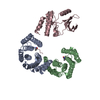 8vyvC  8vywC M: map data used to model this data C: citing same article ( |
|---|---|
| Similar structure data |


-Links
 PDBj
PDBj


- Assembly
| Deposited unit | 
|
|---|---|
| 1 |
|
-Components
| #1: Protein | Mass: 84784.727 Da / Num. of mol.: 1 / Mutation: V600E Source method: isolated from a genetically manipulated source Source: (gene. exp.) Homo sapiens (human) / Cell line: FreeStyle 293-F cells / Gene: BRAF, BRAF1, RAFB1 / Plasmid: pCDNA3.1-FLAG-TEV-BRAF V600E / Cell (production host): FreeStyle 293-F cells / Production host: Homo sapiens (human)References: UniProt: P15056, non-specific serine/threonine protein kinase | ||||
|---|---|---|---|---|---|
| #2: Protein | Mass: 27777.092 Da / Num. of mol.: 2 / Source method: isolated from a natural source / Source: (natural) Homo sapiens (human) / Cell line: FreeStyle 293-F cells / References: UniProt: P63104Has ligand of interest | N | Has protein modification | Y | |
-Experimental details
-Experiment
| Experiment | Method: ELECTRON MICROSCOPY |
|---|---|
| EM experiment | Aggregation state: PARTICLE / 3D reconstruction method: single particle reconstruction |
- Sample preparation
| Component | Name: BRAF V600E - 14-3-3 complex bound to GDC-0879 / Type: COMPLEX / Entity ID: all / Source: MULTIPLE SOURCES | ||||||||||||||||||||||||||||||
|---|---|---|---|---|---|---|---|---|---|---|---|---|---|---|---|---|---|---|---|---|---|---|---|---|---|---|---|---|---|---|---|
| Molecular weight | Value: 0.141 MDa / Experimental value: NO | ||||||||||||||||||||||||||||||
| Source (natural) | Organism: Homo sapiens (human) | ||||||||||||||||||||||||||||||
| Source (recombinant) | Organism: Homo sapiens (human) / Cell: FreeStyle 293-F cells / Plasmid: pCDNA3.1-FLAG-TEV-BRAF V600E | ||||||||||||||||||||||||||||||
| Buffer solution | pH: 7.5 | ||||||||||||||||||||||||||||||
| Buffer component |
| ||||||||||||||||||||||||||||||
| Specimen | Conc.: 1 mg/ml / Embedding applied: NO / Shadowing applied: NO / Staining applied: NO / Vitrification applied: YES | ||||||||||||||||||||||||||||||
| Vitrification | Cryogen name: ETHANE / Details: Chameleon system (SPT Labtech) |
- Electron microscopy imaging
| Experimental equipment |  Model: Titan Krios / Image courtesy: FEI Company |
|---|---|
| Microscopy | Model: TFS KRIOS |
| Electron gun | Electron source:  FIELD EMISSION GUN / Accelerating voltage: 300 kV / Illumination mode: FLOOD BEAM FIELD EMISSION GUN / Accelerating voltage: 300 kV / Illumination mode: FLOOD BEAM |
| Electron lens | Mode: BRIGHT FIELD / Nominal defocus max: 4600 nm / Nominal defocus min: 1700 nm |
| Image recording | Electron dose: 51.55 e/Å2 / Film or detector model: GATAN K3 BIOQUANTUM (6k x 4k) |
- Processing
| EM software |
| ||||||||||||||||||||||||
|---|---|---|---|---|---|---|---|---|---|---|---|---|---|---|---|---|---|---|---|---|---|---|---|---|---|
| CTF correction | Type: PHASE FLIPPING AND AMPLITUDE CORRECTION | ||||||||||||||||||||||||
| 3D reconstruction | Resolution: 4.32 Å / Resolution method: FSC 0.143 CUT-OFF / Num. of particles: 32966 Details: Resolution corresponds to the FSC 0.143 cut-off of the masked map Symmetry type: POINT | ||||||||||||||||||||||||
| Refinement | Cross valid method: NONE Stereochemistry target values: GeoStd + Monomer Library + CDL v1.2 | ||||||||||||||||||||||||
| Displacement parameters | Biso mean: 165.84 Å2 | ||||||||||||||||||||||||
| Refine LS restraints |
|
