 Movie
Movie Controller

[English] 日本語
 Yorodumi
- PDB-8vx9: Structure of HamAB apo complex from the Escherichia coli Hachiman... -
+ Open data

- Basic information
| Entry | Database: PDB / ID: 8vx9 | ||||||
|---|---|---|---|---|---|---|---|
| Title | Structure of HamAB apo complex from the Escherichia coli Hachiman defense system | ||||||
 Components Components |
| ||||||
 Keywords Keywords | IMMUNE SYSTEM / Nuclease-helicase complex / antiphage defense system | ||||||
| Function / homology |  Function and homology information Function and homology information | ||||||
| Biological species |  | ||||||
| Method | ELECTRON MICROSCOPY / single particle reconstruction / cryo EM / Resolution: 2.65 Å | ||||||
 Authors Authors | Tuck, O.T. / Doudna, J.A. | ||||||
| Funding support |  United States, 1items United States, 1items
| ||||||
 Citation Citation | Journal: bioRxiv / Year: 2024 Title: Hachiman is a genome integrity sensor. Authors: Owen T Tuck / Benjamin A Adler / Emily G Armbruster / Arushi Lahiri / Jason J Hu / Julia Zhou / Joe Pogliano / Jennifer A Doudna / Abstract: Hachiman is a broad-spectrum antiphage defense system of unknown function. We show here that Hachiman comprises a heterodimeric nuclease-helicase complex, HamAB. HamA, previously a protein of unknown ...Hachiman is a broad-spectrum antiphage defense system of unknown function. We show here that Hachiman comprises a heterodimeric nuclease-helicase complex, HamAB. HamA, previously a protein of unknown function, is the effector nuclease. HamB is the sensor helicase. HamB constrains HamA activity during surveillance of intact dsDNA. When the HamAB complex detects DNA damage, HamB helicase activity liberates HamA, unleashing nuclease activity. Hachiman activation degrades all DNA in the cell, creating 'phantom' cells devoid of both phage and host DNA. We demonstrate Hachiman activation in the absence of phage by treatment with DNA-damaging agents, suggesting that Hachiman responds to aberrant DNA states. Phylogenetic similarities between the Hachiman helicase and eukaryotic enzymes suggest this bacterial immune system has been repurposed for diverse functions across all domains of life. | ||||||
| History |
|
- Structure visualization
| Structure viewer | Molecule: MolmilJmol/JSmol |
|---|
- Downloads & links
-Download
| PDBx/mmCIF format | 8vx9.cif.gz | 282.5 KB | Display | PDBx/mmCIF format |
|---|---|---|---|---|
| PDB format | pdb8vx9.ent.gz | 224.9 KB | Display | PDB format |
| PDBx/mmJSON format | 8vx9.json.gz | Tree view | PDBx/mmJSON format | |
| Others |  Other downloads Other downloads |
-Validation report
| Arichive directory | https://data.pdbj.org/pub/pdb/validation_reports/vx/8vx9ftp://data.pdbj.org/pub/pdb/validation_reports/vx/8vx9 | HTTPS FTP |
|---|
-Related structure data
| Related structure data | 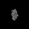 43613MC  8vxaC  8vxcC  8vxyC M: map data used to model this data C: citing same article ( |
|---|---|
| Similar structure data |

-Links
 PDBj
PDBj- Assembly
| Deposited unit | 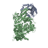
|
|---|---|
| 1 |
|
-Components
| #1: Protein | Mass: 28923.797 Da / Num. of mol.: 1 Source method: isolated from a genetically manipulated source Source: (gene. exp.) |
|---|---|
| #2: Protein | Mass: 130898.453 Da / Num. of mol.: 1 Source method: isolated from a genetically manipulated source Source: (gene. exp.) |
-Experimental details
-Experiment
| Experiment | Method: ELECTRON MICROSCOPY |
|---|---|
| EM experiment | Aggregation state: PARTICLE / 3D reconstruction method: single particle reconstruction |
- Sample preparation
| Component | Name: Apo HamAB complex / Type: COMPLEX / Entity ID: all / Source: RECOMBINANT | |||||||||||||||||||||||||
|---|---|---|---|---|---|---|---|---|---|---|---|---|---|---|---|---|---|---|---|---|---|---|---|---|---|---|
| Molecular weight | Value: .16 MDa / Experimental value: NO | |||||||||||||||||||||||||
| Source (natural) | Organism: | |||||||||||||||||||||||||
| Source (recombinant) | Organism: | |||||||||||||||||||||||||
| Buffer solution | pH: 8 | |||||||||||||||||||||||||
| Buffer component |
| |||||||||||||||||||||||||
| Specimen | Conc.: 1 mg/ml / Embedding applied: NO / Shadowing applied: NO / Staining applied: NO / Vitrification applied: YES | |||||||||||||||||||||||||
| Specimen support | Grid material: GOLD / Grid mesh size: 200 divisions/in. / Grid type: UltrAuFoil R2/2 | |||||||||||||||||||||||||
| Vitrification | Instrument: FEI VITROBOT MARK IV / Cryogen name: ETHANE / Humidity: 100 % / Chamber temperature: 281 K |
- Electron microscopy imaging
| Experimental equipment |  Model: Titan Krios / Image courtesy: FEI Company |
|---|---|
| Microscopy | Model: FEI TITAN KRIOS |
| Electron gun | Electron source:  FIELD EMISSION GUN / Accelerating voltage: 300 kV / Illumination mode: OTHER FIELD EMISSION GUN / Accelerating voltage: 300 kV / Illumination mode: OTHER |
| Electron lens | Mode: BRIGHT FIELD / Nominal defocus max: 1800 nm / Nominal defocus min: 800 nm / Cs: 2.7 mm / Alignment procedure: COMA FREE |
| Specimen holder | Cryogen: NITROGEN |
| Image recording | Electron dose: 50 e/Å2 / Film or detector model: GATAN K3 BIOQUANTUM (6k x 4k) / Num. of grids imaged: 1 / Num. of real images: 4796 |
- Processing
| EM software |
| ||||||||||||||||||||||||||||||||||||||||||||||||||
|---|---|---|---|---|---|---|---|---|---|---|---|---|---|---|---|---|---|---|---|---|---|---|---|---|---|---|---|---|---|---|---|---|---|---|---|---|---|---|---|---|---|---|---|---|---|---|---|---|---|---|---|
| CTF correction | Type: PHASE FLIPPING AND AMPLITUDE CORRECTION | ||||||||||||||||||||||||||||||||||||||||||||||||||
| Particle selection | Num. of particles selected: 5538550 | ||||||||||||||||||||||||||||||||||||||||||||||||||
| 3D reconstruction | Resolution: 2.65 Å / Resolution method: FSC 0.143 CUT-OFF / Num. of particles: 309630 / Symmetry type: POINT | ||||||||||||||||||||||||||||||||||||||||||||||||||
| Atomic model building | Protocol: OTHER / Space: REAL | ||||||||||||||||||||||||||||||||||||||||||||||||||
| Atomic model building | Details: v1.4.0 / Source name: AlphaFold / Type: in silico model |
