[English] 日本語
 Yorodumi
- PDB-8vu3: Cryo-EM structure of cyanobacterial PSI with bound platinum nanop... -
+ Open data
- Basic information
| Entry | Database: PDB / ID: 8vu3 | ||||||||||||
|---|---|---|---|---|---|---|---|---|---|---|---|---|---|
| Title | Cryo-EM structure of cyanobacterial PSI with bound platinum nanoparticles | ||||||||||||
 Components Components |
| ||||||||||||
 Keywords Keywords | PHOTOSYNTHESIS / cyanobacteria / biohybrid / platinum nanoparticles / hydrogen production / photosystem I / solar fuel | ||||||||||||
| Function / homology |  Function and homology information Function and homology informationphotosystem I reaction center / photosystem I / photosynthetic electron transport in photosystem I / photosystem I / plasma membrane-derived thylakoid membrane / chlorophyll binding / photosynthesis / endomembrane system / 4 iron, 4 sulfur cluster binding / electron transfer activity ...photosystem I reaction center / photosystem I / photosynthetic electron transport in photosystem I / photosystem I / plasma membrane-derived thylakoid membrane / chlorophyll binding / photosynthesis / endomembrane system / 4 iron, 4 sulfur cluster binding / electron transfer activity / oxidoreductase activity / magnesium ion binding / metal ion binding / membrane Similarity search - Function | ||||||||||||
| Biological species |  Thermostichus lividus (bacteria) Thermostichus lividus (bacteria) | ||||||||||||
| Method | ELECTRON MICROSCOPY / single particle reconstruction / cryo EM / Resolution: 2.27 Å | ||||||||||||
 Authors Authors | Gisriel, C.J. / Malavath, T. / Brudvig, G.W. / Utschig, L.M. | ||||||||||||
| Funding support |  United States, 3items United States, 3items
| ||||||||||||
 Citation Citation |  Journal: Nat Commun / Year: 2024 Journal: Nat Commun / Year: 2024Title: Structure of a biohybrid photosystem I-platinum nanoparticle solar fuel catalyst. Authors: Christopher J Gisriel / Tirupathi Malavath / Tianyin Qiu / Jan Paul Menzel / Victor S Batista / Gary W Brudvig / Lisa M Utschig /  Abstract: Biohybrid solar fuel catalysts leverage natural light-driven enzymes to produce valuable fuel products. One useful biological platform for such a system is photosystem I, a pigment-protein complex ...Biohybrid solar fuel catalysts leverage natural light-driven enzymes to produce valuable fuel products. One useful biological platform for such a system is photosystem I, a pigment-protein complex that captures sunlight and converts it into chemical energy with near unity quantum efficiency, which generates low potential reducing equivalents for metabolism. Realizing and understanding the molecular basis for an approach that utilizes those electrons and stores solar energy as a fuel is therefore appealing. Here, we report the 2.27-Å global resolution cryo-EM structure of a photosystem I complex with bound platinum nanoparticles that catalyzes light-driven H production. The platinum nanoparticle binding sites and possible stabilizing interactions are described. Overall, the investigation reveals a direct structural look at a photon-to-fuels photosynthetic biohybrid system. | ||||||||||||
| History |
|
- Structure visualization
| Structure viewer | Molecule:  Molmil Molmil Jmol/JSmol Jmol/JSmol |
|---|
- Downloads & links
- Download
| PDBx/mmCIF format |  8vu3.cif.gz 8vu3.cif.gz | 1.7 MB | Display |  PDBx/mmCIF format PDBx/mmCIF format |
|---|---|---|---|---|
| PDB format |  pdb8vu3.ent.gz pdb8vu3.ent.gz | 1.5 MB | Display |  PDB format PDB format |
| PDBx/mmJSON format |  8vu3.json.gz 8vu3.json.gz | Tree view |  PDBx/mmJSON format PDBx/mmJSON format | |
| Others |  Other downloads Other downloads |
-Validation report
| Arichive directory |  https://data.pdbj.org/pub/pdb/validation_reports/vu/8vu3 https://data.pdbj.org/pub/pdb/validation_reports/vu/8vu3 ftp://data.pdbj.org/pub/pdb/validation_reports/vu/8vu3 ftp://data.pdbj.org/pub/pdb/validation_reports/vu/8vu3 | HTTPS FTP |
|---|
-Related structure data
| Related structure data |  43525MC M: map data used to model this data C: citing same article ( |
|---|---|
| Similar structure data | Similarity search - Function & homology  F&H Search F&H Search |
- Links
Links
- Assembly
| Deposited unit | 
|
|---|---|
| 1 |
|
- Components
-Photosystem I P700 chlorophyll a apoprotein ... , 2 types, 6 molecules AGaBHb
| #1: Protein | Mass: 83195.555 Da / Num. of mol.: 3 / Source method: isolated from a natural source / Source: (natural)  Thermostichus lividus (bacteria) / References: UniProt: A0A2D2Q201, photosystem I Thermostichus lividus (bacteria) / References: UniProt: A0A2D2Q201, photosystem I#2: Protein | Mass: 83152.586 Da / Num. of mol.: 3 / Source method: isolated from a natural source / Source: (natural)  Thermostichus lividus (bacteria) / References: UniProt: A0A2D2Q1Z9, photosystem I Thermostichus lividus (bacteria) / References: UniProt: A0A2D2Q1Z9, photosystem I |
|---|
-Photosystem I reaction center subunit ... , 8 types, 24 molecules DOdEPeFQfIRiJSjKTkLUlMVm
| #4: Protein | Mass: 15408.582 Da / Num. of mol.: 3 / Source method: isolated from a natural source / Source: (natural)  Thermostichus lividus (bacteria) / References: UniProt: A0A2D2Q2Y6 Thermostichus lividus (bacteria) / References: UniProt: A0A2D2Q2Y6#5: Protein | Mass: 8238.266 Da / Num. of mol.: 3 / Source method: isolated from a natural source / Source: (natural)  Thermostichus lividus (bacteria) / References: UniProt: A0A2D2Q1I4 Thermostichus lividus (bacteria) / References: UniProt: A0A2D2Q1I4#6: Protein | Mass: 17654.516 Da / Num. of mol.: 3 / Source method: isolated from a natural source / Source: (natural)  Thermostichus lividus (bacteria) / References: UniProt: A0A2D2PYV7 Thermostichus lividus (bacteria) / References: UniProt: A0A2D2PYV7#7: Protein/peptide | Mass: 4480.442 Da / Num. of mol.: 3 / Source method: isolated from a natural source / Source: (natural)  Thermostichus lividus (bacteria) / References: UniProt: A0A2D2Q0W5 Thermostichus lividus (bacteria) / References: UniProt: A0A2D2Q0W5#8: Protein/peptide | Mass: 4965.914 Da / Num. of mol.: 3 / Source method: isolated from a natural source / Source: (natural)  Thermostichus lividus (bacteria) / References: UniProt: A0A2D2PZG9 Thermostichus lividus (bacteria) / References: UniProt: A0A2D2PZG9#9: Protein | Mass: 8471.931 Da / Num. of mol.: 3 / Source method: isolated from a natural source / Source: (natural)  Thermostichus lividus (bacteria) / References: UniProt: A0A2D2Q0K8 Thermostichus lividus (bacteria) / References: UniProt: A0A2D2Q0K8#10: Protein | Mass: 16233.698 Da / Num. of mol.: 3 / Source method: isolated from a natural source / Source: (natural)  Thermostichus lividus (bacteria) / References: UniProt: A0A2D2Q0R7 Thermostichus lividus (bacteria) / References: UniProt: A0A2D2Q0R7#11: Protein/peptide | Mass: 3410.116 Da / Num. of mol.: 3 / Source method: isolated from a natural source / Source: (natural)  Thermostichus lividus (bacteria) / References: UniProt: A0A3B7MIX8 Thermostichus lividus (bacteria) / References: UniProt: A0A3B7MIX8 |
|---|
-Protein / Protein/peptide , 2 types, 6 molecules CNcWXx
| #12: Protein/peptide | Mass: 4134.966 Da / Num. of mol.: 3 / Source method: isolated from a natural source / Source: (natural)  Thermostichus lividus (bacteria) / References: UniProt: A0A2D2Q2C6 Thermostichus lividus (bacteria) / References: UniProt: A0A2D2Q2C6#3: Protein | Mass: 8809.207 Da / Num. of mol.: 3 / Source method: isolated from a natural source / Source: (natural)  Thermostichus lividus (bacteria) / References: UniProt: A0A2D2Q1F9, photosystem I Thermostichus lividus (bacteria) / References: UniProt: A0A2D2Q1F9, photosystem I |
|---|
-Non-polymers , 8 types, 381 molecules 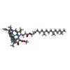
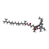
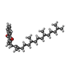

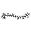



| #13: Chemical | | #14: Chemical | ChemComp-CLA / #15: Chemical | ChemComp-PQN / #16: Chemical | ChemComp-SF4 / #17: Chemical | ChemComp-BCR / #18: Chemical | ChemComp-LHG / #19: Chemical | #20: Chemical | |
|---|
-Details
| Has ligand of interest | Y |
|---|---|
| Has protein modification | Y |
-Experimental details
-Experiment
| Experiment | Method: ELECTRON MICROSCOPY |
|---|---|
| EM experiment | Aggregation state: PARTICLE / 3D reconstruction method: single particle reconstruction |
- Sample preparation
| Component | Name: Photosystem I from Thermostichus lividus / Type: COMPLEX / Entity ID: #1-#12 / Source: NATURAL |
|---|---|
| Source (natural) | Organism:  Thermostichus lividus (bacteria) Thermostichus lividus (bacteria) |
| Buffer solution | pH: 8 |
| Specimen | Conc.: 1 mg/ml / Embedding applied: NO / Shadowing applied: NO / Staining applied: NO / Vitrification applied: YES |
| Vitrification | Cryogen name: ETHANE |
- Electron microscopy imaging
| Experimental equipment |  Model: Titan Krios / Image courtesy: FEI Company |
|---|---|
| Microscopy | Model: FEI TITAN KRIOS |
| Electron gun | Electron source:  FIELD EMISSION GUN / Accelerating voltage: 300 kV / Illumination mode: FLOOD BEAM FIELD EMISSION GUN / Accelerating voltage: 300 kV / Illumination mode: FLOOD BEAM |
| Electron lens | Mode: BRIGHT FIELD / Nominal defocus max: 2200 nm / Nominal defocus min: 800 nm |
| Image recording | Electron dose: 40 e/Å2 / Film or detector model: GATAN K3 (6k x 4k) |
- Processing
| EM software | Name: PHENIX / Version: 1.20.1_4487: / Category: model refinement | ||||||||||||||||||||||||
|---|---|---|---|---|---|---|---|---|---|---|---|---|---|---|---|---|---|---|---|---|---|---|---|---|---|
| CTF correction | Type: PHASE FLIPPING AND AMPLITUDE CORRECTION | ||||||||||||||||||||||||
| 3D reconstruction | Resolution: 2.27 Å / Resolution method: FSC 0.143 CUT-OFF / Num. of particles: 189655 / Symmetry type: POINT | ||||||||||||||||||||||||
| Refine LS restraints |
|
 Movie
Movie Controller


 PDBj
PDBj


