 Movie
Movie Controller

[English] 日本語
 Yorodumi
- PDB-8vqh: CryoEM structure of BchN-BchB electron acceptor component protein... -
+ Open data

- Basic information
| Entry | Database: PDB / ID: 8vqh | ||||||||||||||||||||||||
|---|---|---|---|---|---|---|---|---|---|---|---|---|---|---|---|---|---|---|---|---|---|---|---|---|---|
| Title | CryoEM structure of BchN-BchB electron acceptor component protein of DPOR | ||||||||||||||||||||||||
 Components Components |
| ||||||||||||||||||||||||
 Keywords Keywords | OXIDOREDUCTASE / Plant Protein / Electron Transfer Enzymes / Photosynthesis | ||||||||||||||||||||||||
| Function / homology |  Function and homology information Function and homology informationferredoxin:protochlorophyllide reductase (ATP-dependent) / photosynthesis, dark reaction / light-independent bacteriochlorophyll biosynthetic process / oxidoreductase activity, acting on iron-sulfur proteins as donors / oxidoreductase activity, acting on the CH-CH group of donors, iron-sulfur protein as acceptor / SUMO is conjugated to E1 (UBA2:SAE1) / SUMOylation of nuclear envelope proteins / SUMO is transferred from E1 to E2 (UBE2I, UBC9) / SUMO is proteolytically processed / SUMOylation of transcription factors ...ferredoxin:protochlorophyllide reductase (ATP-dependent) / photosynthesis, dark reaction / light-independent bacteriochlorophyll biosynthetic process / oxidoreductase activity, acting on iron-sulfur proteins as donors / oxidoreductase activity, acting on the CH-CH group of donors, iron-sulfur protein as acceptor / SUMO is conjugated to E1 (UBA2:SAE1) / SUMOylation of nuclear envelope proteins / SUMO is transferred from E1 to E2 (UBE2I, UBC9) / SUMO is proteolytically processed / SUMOylation of transcription factors / Postmitotic nuclear pore complex (NPC) reformation / SUMOylation of transcription cofactors / septin ring / SUMOylation of DNA damage response and repair proteins / Transcriptional and post-translational regulation of MITF-M expression and activity / SUMOylation of DNA replication proteins / SUMOylation of SUMOylation proteins / Recruitment and ATM-mediated phosphorylation of repair and signaling proteins at DNA double strand breaks / SUMOylation of RNA binding proteins / SUMOylation of chromatin organization proteins / ubiquitin-like protein ligase binding / protein sumoylation / condensed nuclear chromosome / protein tag activity / 4 iron, 4 sulfur cluster binding / ATP binding / metal ion binding / identical protein binding / nucleus Similarity search - Function | ||||||||||||||||||||||||
| Biological species |  Cereibacter sphaeroides (bacteria) Cereibacter sphaeroides (bacteria) | ||||||||||||||||||||||||
| Method | ELECTRON MICROSCOPY / single particle reconstruction / cryo EM / Resolution: 2.7 Å | ||||||||||||||||||||||||
 Authors Authors | Kashyap, R. / Antony, E. | ||||||||||||||||||||||||
| Funding support |  United States, 1items United States, 1items
| ||||||||||||||||||||||||
 Citation Citation | Journal: Nat Commun / Year: 2025 Title: Cryo-EM captures the coordination of asymmetric electron transfer through a di-copper site in DPOR. Authors: Rajnandani Kashyap / Natalie Walsh / Jaigeeth Deveryshetty / Monika Tokmina-Lukaszewska / Kewei Zhao / Yunqiao J Gan / Brian M Hoffman / Ritimukta Sarangi / Brian Bothner / Brian Bennett / Edwin Antony / Abstract: Enzymes that catalyze long-range electron transfer (ET) reactions often function as higher order complexes that possess two structurally symmetrical halves. The functional advantages for such an ...Enzymes that catalyze long-range electron transfer (ET) reactions often function as higher order complexes that possess two structurally symmetrical halves. The functional advantages for such an architecture remain a mystery. Using cryoelectron microscopy we capture snapshots of the nitrogenase-like dark-operative protochlorophyllide oxidoreductase (DPOR) during substrate binding and turnover. DPOR catalyzes reduction of the C17 = C18 double bond in protochlorophyllide during the dark chlorophyll biosynthetic pathway. DPOR is composed of electron donor (L-protein) and acceptor (NB-protein) component proteins that transiently form a complex in the presence of ATP to facilitate ET. NB-protein is an αβ heterotetramer with two structurally identical halves. However, our structures reveal that NB-protein becomes functionally asymmetric upon substrate binding. Asymmetry results in allosteric inhibition of L-protein engagement and ET in one half. Residues that form a conduit for ET are aligned in one half while misaligned in the other. An ATP hydrolysis-coupled conformational switch is triggered once ET is accomplished in one half. These structural changes are then relayed to the other half through a di-nuclear copper center at the tetrameric interface of the NB-protein and leads to activation of ET and substrate reduction. These findings provide a mechanistic blueprint for regulation of long-range electron transfer reactions. | ||||||||||||||||||||||||
| History |
|
- Structure visualization
| Structure viewer | Molecule: MolmilJmol/JSmol |
|---|
- Downloads & links
-Download
| PDBx/mmCIF format | 8vqh.cif.gz | 327.2 KB | Display | PDBx/mmCIF format |
|---|---|---|---|---|
| PDB format | pdb8vqh.ent.gz | 256.3 KB | Display | PDB format |
| PDBx/mmJSON format | 8vqh.json.gz | Tree view | PDBx/mmJSON format | |
| Others |  Other downloads Other downloads |
-Validation report
| Arichive directory | https://data.pdbj.org/pub/pdb/validation_reports/vq/8vqhftp://data.pdbj.org/pub/pdb/validation_reports/vq/8vqh | HTTPS FTP |
|---|
-Related structure data
| Related structure data | 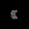 43443MC 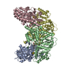 8vqiC 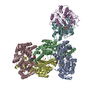 8vqjC 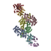 9buoC 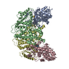 9e7hC 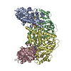 9efuC M: map data used to model this data C: citing same article ( |
|---|---|
| Similar structure data |

-Links
 PDBj
PDBj


- Assembly
| Deposited unit | 
|
|---|---|
| 1 |
|
-Components
| #1: Protein | Mass: 46188.773 Da / Num. of mol.: 2 Source method: isolated from a genetically manipulated source Source: (gene. exp.) Cereibacter sphaeroides (bacteria) / Gene: bchN, RSKD131_1611 / Production host: References: UniProt: B9KK24, ferredoxin:protochlorophyllide reductase (ATP-dependent) #2: Protein | Mass: 69448.922 Da / Num. of mol.: 2 Source method: isolated from a genetically manipulated source Source: (gene. exp.) Cereibacter sphaeroides (bacteria) / Gene: SMT3, YDR510W, D9719.15, bchB, RHOS4_18910, RSP_0286 / Production host: References: UniProt: Q12306, UniProt: Q9Z5D9, ferredoxin:protochlorophyllide reductase (ATP-dependent) #3: Chemical |   Mass: 351.640 Da / Num. of mol.: 2 / Source method: obtained synthetically / Formula: Fe4S4 / Feature type: SUBJECT OF INVESTIGATION Mass: 351.640 Da / Num. of mol.: 2 / Source method: obtained synthetically / Formula: Fe4S4 / Feature type: SUBJECT OF INVESTIGATION#4: Chemical |   Mass: 63.546 Da / Num. of mol.: 2 / Source method: obtained synthetically / Formula: Cu / Feature type: SUBJECT OF INVESTIGATION Mass: 63.546 Da / Num. of mol.: 2 / Source method: obtained synthetically / Formula: Cu / Feature type: SUBJECT OF INVESTIGATION#5: Water | ChemComp-HOH / |  Mass: 18.015 Da / Num. of mol.: 2 / Source method: isolated from a natural source / Formula: H2O Mass: 18.015 Da / Num. of mol.: 2 / Source method: isolated from a natural source / Formula: H2OHas ligand of interest | Y | Has protein modification | N | |
|---|
-Experimental details
-Experiment
| Experiment | Method: ELECTRON MICROSCOPY |
|---|---|
| EM experiment | Aggregation state: PARTICLE / 3D reconstruction method: single particle reconstruction |
- Sample preparation
| Component | Name: CryoEM structure of BchN-BchB electron acceptor component protein of DPOR Type: COMPLEX / Entity ID: #1-#2 / Source: RECOMBINANT |
|---|---|
| Molecular weight | Value: 0.236 MDa / Experimental value: NO |
| Source (natural) | Organism: Cereibacter sphaeroides (bacteria) |
| Source (recombinant) | Organism: |
| Buffer solution | pH: 7.5 |
| Specimen | Embedding applied: NO / Shadowing applied: NO / Staining applied: NO / Vitrification applied: YES |
| Vitrification | Instrument: FEI VITROBOT MARK IV / Cryogen name: ETHANE / Humidity: 100 % / Chamber temperature: 277.15 K |
- Electron microscopy imaging
| Experimental equipment |  Model: Titan Krios / Image courtesy: FEI Company |
|---|---|
| Microscopy | Model: TFS KRIOS |
| Electron gun | Electron source:  FIELD EMISSION GUN / Accelerating voltage: 300 kV / Illumination mode: OTHER FIELD EMISSION GUN / Accelerating voltage: 300 kV / Illumination mode: OTHER |
| Electron lens | Mode: BRIGHT FIELD / Nominal magnification: 105000 X / Nominal defocus max: 2200 nm / Nominal defocus min: 1000 nm / Cs: 2.7 mm / C2 aperture diameter: 70 µm |
| Image recording | Electron dose: 50 e/Å2 / Film or detector model: GATAN K3 BIOQUANTUM (6k x 4k) |
- Processing
| CTF correction | Type: NONE | ||||||||||||||||||||||||
|---|---|---|---|---|---|---|---|---|---|---|---|---|---|---|---|---|---|---|---|---|---|---|---|---|---|
| Symmetry | Point symmetry: C1 (asymmetric) | ||||||||||||||||||||||||
| 3D reconstruction | Resolution: 2.7 Å / Resolution method: FSC 0.143 CUT-OFF / Num. of particles: 160048 / Symmetry type: POINT | ||||||||||||||||||||||||
| Refinement | Highest resolution: 2.7 Å Stereochemistry target values: REAL-SPACE (WEIGHTED MAP SUM AT ATOM CENTERS) | ||||||||||||||||||||||||
| Refine LS restraints |
|
