 Movie
Movie Controller


+ Open data

- Basic information
| Entry | 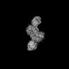 | |||||||||
|---|---|---|---|---|---|---|---|---|---|---|
| Title | CryoEM structure of DPOR in the presence of ADP-AlF3 | |||||||||
 Map data Map data | ||||||||||
 Sample Sample |
| |||||||||
 Keywords Keywords | Plant Protein / Electron Transfer Enzymes / Photosynthesis / OXIDOREDUCTASE | |||||||||
| Function / homology |  Function and homology information Function and homology informationferredoxin:protochlorophyllide reductase (ATP-dependent) / photosynthesis, dark reaction / light-independent bacteriochlorophyll biosynthetic process / oxidoreductase activity, acting on iron-sulfur proteins as donors / oxidoreductase activity, acting on the CH-CH group of donors, iron-sulfur protein as acceptor / 4 iron, 4 sulfur cluster binding / ATP binding / metal ion binding Similarity search - Function | |||||||||
| Biological species |  Cereibacter sphaeroides (bacteria) Cereibacter sphaeroides (bacteria) | |||||||||
| Method | single particle reconstruction / cryo EM / Resolution: 3.68 Å | |||||||||
 Authors Authors | Kashyap R / Antony E | |||||||||
| Funding support |  United States, 1 items United States, 1 items
| |||||||||
 Citation Citation | Journal: Nat Commun / Year: 2025 Title: Cryo-EM captures the coordination of asymmetric electron transfer through a di-copper site in DPOR. Authors: Rajnandani Kashyap / Natalie Walsh / Jaigeeth Deveryshetty / Monika Tokmina-Lukaszewska / Kewei Zhao / Yunqiao J Gan / Brian M Hoffman / Ritimukta Sarangi / Brian Bothner / Brian Bennett / Edwin Antony / Abstract: Enzymes that catalyze long-range electron transfer (ET) reactions often function as higher order complexes that possess two structurally symmetrical halves. The functional advantages for such an ...Enzymes that catalyze long-range electron transfer (ET) reactions often function as higher order complexes that possess two structurally symmetrical halves. The functional advantages for such an architecture remain a mystery. Using cryoelectron microscopy we capture snapshots of the nitrogenase-like dark-operative protochlorophyllide oxidoreductase (DPOR) during substrate binding and turnover. DPOR catalyzes reduction of the C17 = C18 double bond in protochlorophyllide during the dark chlorophyll biosynthetic pathway. DPOR is composed of electron donor (L-protein) and acceptor (NB-protein) component proteins that transiently form a complex in the presence of ATP to facilitate ET. NB-protein is an αβ heterotetramer with two structurally identical halves. However, our structures reveal that NB-protein becomes functionally asymmetric upon substrate binding. Asymmetry results in allosteric inhibition of L-protein engagement and ET in one half. Residues that form a conduit for ET are aligned in one half while misaligned in the other. An ATP hydrolysis-coupled conformational switch is triggered once ET is accomplished in one half. These structural changes are then relayed to the other half through a di-nuclear copper center at the tetrameric interface of the NB-protein and leads to activation of ET and substrate reduction. These findings provide a mechanistic blueprint for regulation of long-range electron transfer reactions. | |||||||||
| History |
|
- Structure visualization
| Supplemental images |
|---|

- Downloads & links
-EMDB archive
| Map data | emd_44913.map.gz | 398.7 MB | EMDB map data format | |
|---|---|---|---|---|
| Header (meta data) | emd-44913-v30.xmlemd-44913.xml | 18.9 KB 18.9 KB | Display Display | EMDB header |
| FSC (resolution estimation) | emd_44913_fsc.xml | 15.9 KB | Display | FSC data file |
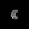
| Images |  emd_44913.png emd_44913.png | 51 KB | ||
| Filedesc metadata | emd-44913.cif.gz | 6.6 KB | ||
| Others | emd_44913_half_map_1.map.gzemd_44913_half_map_2.map.gz | 391.3 MB 391.3 MB | ||
| Archive directory |  http://ftp.pdbj.org/pub/emdb/structures/EMD-44913ftp://ftp.pdbj.org/pub/emdb/structures/EMD-44913 http://ftp.pdbj.org/pub/emdb/structures/EMD-44913ftp://ftp.pdbj.org/pub/emdb/structures/EMD-44913 | HTTPS FTP |
-Related structure data
| Related structure data | 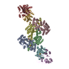 9buoMC 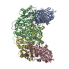 8vqhC 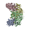 8vqiC 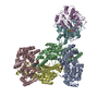 8vqjC 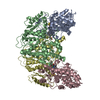 9e7hC 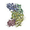 9efuC M: atomic model generated by this map C: citing same article ( |
|---|---|
| Similar structure data |

-Links
| EMDB pages | EMDB (EBI/PDBe) / EMDataResource |
|---|---|
| Related items in Molecule of the Month |

-Map
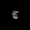
| File | Download / File: emd_44913.map.gz / Format: CCP4 / Size: 421.9 MB / Type: IMAGE STORED AS FLOATING POINT NUMBER (4 BYTES) | ||||||||||||||||||||||||||||||||||||
|---|---|---|---|---|---|---|---|---|---|---|---|---|---|---|---|---|---|---|---|---|---|---|---|---|---|---|---|---|---|---|---|---|---|---|---|---|---|
| Projections & slices | Image control
Images are generated by Spider. | ||||||||||||||||||||||||||||||||||||
| Voxel size | X=Y=Z: 0.82 Å | ||||||||||||||||||||||||||||||||||||
| Density |
| ||||||||||||||||||||||||||||||||||||
| Symmetry | Space group: 1 | ||||||||||||||||||||||||||||||||||||
| Details | EMDB XML:
|
 Z (Sec.)
 Y (Row.)
 X (Col.)


-Supplemental data
-Half map: #2
| File | emd_44913_half_map_1.map | ||||||||||||
|---|---|---|---|---|---|---|---|---|---|---|---|---|---|
| Projections & Slices |
| ||||||||||||
| Density Histograms |


-Half map: #1
| File | emd_44913_half_map_2.map | ||||||||||||
|---|---|---|---|---|---|---|---|---|---|---|---|---|---|
| Projections & Slices |
| ||||||||||||
| Density Histograms |


- Sample components
-Entire : CryoEM structure of DPOR in the presence of ADP-AlF3
| Entire | Name: CryoEM structure of DPOR in the presence of ADP-AlF3 |
|---|---|
| Components |
|
-Supramolecule #1: CryoEM structure of DPOR in the presence of ADP-AlF3
| Supramolecule | Name: CryoEM structure of DPOR in the presence of ADP-AlF3 / type: complex / ID: 1 / Parent: 0 / Macromolecule list: #1-#3 |
|---|---|
| Source (natural) | Organism: Cereibacter sphaeroides (bacteria) |
| Molecular weight | Theoretical: 347.8 KDa |
-Macromolecule #1: Light-independent protochlorophyllide reductase iron-sulfur ATP-b...
| Macromolecule | Name: Light-independent protochlorophyllide reductase iron-sulfur ATP-binding protein type: protein_or_peptide / ID: 1 / Number of copies: 4 / Enantiomer: LEVO EC number: ferredoxin:protochlorophyllide reductase (ATP-dependent) |
|---|---|
| Source (natural) | Organism: Cereibacter sphaeroides (bacteria) |
| Molecular weight | Theoretical: 34.85034 KDa |
| Recombinant expression | Organism: |
| Sequence | String: MGSSHHHHHH SQDPENLYFQ SMSPKDLTIP TGADGEGSVQ VHLDEADKIT GAKVFAVYGK GGIGKSTTSS NLSAAFSILG KRVLQIGCD PKHDSTFTLT GSLVPTVIDV LKDVDFHPEE LRPEDFVFEG FNGVMCVEAG GPPAGTGCGG YVVGQTVKLL K QHHLLDDT ...String: MGSSHHHHHH SQDPENLYFQ SMSPKDLTIP TGADGEGSVQ VHLDEADKIT GAKVFAVYGK GGIGKSTTSS NLSAAFSILG KRVLQIGCD PKHDSTFTLT GSLVPTVIDV LKDVDFHPEE LRPEDFVFEG FNGVMCVEAG GPPAGTGCGG YVVGQTVKLL K QHHLLDDT DVVIFDVLGD VVCGGFAAPL QHADQAVVVT ANDFDSIYAM NRIIAAVQAK SKNYKVRLAG CVANRSRATD EV DRFCKET NFRRLAHMPD LDAIRRSRLK KKTLFEMDED QDVLAARAEY IRLAESLWRG LDPIDPHSLP DREIFELLGF D UniProtKB: Light-independent protochlorophyllide reductase iron-sulfur ATP-binding protein |
-Macromolecule #2: Light-independent protochlorophyllide reductase subunit N
| Macromolecule | Name: Light-independent protochlorophyllide reductase subunit N type: protein_or_peptide / ID: 2 / Number of copies: 2 / Enantiomer: LEVO EC number: ferredoxin:protochlorophyllide reductase (ATP-dependent) |
|---|---|
| Source (natural) | Organism: Cereibacter sphaeroides (bacteria) |
| Molecular weight | Theoretical: 46.188773 KDa |
| Recombinant expression | Organism: |
| Sequence | String: MSLDLPPPPA RGCRSTEVLK ERGQREVFCG LTGIIWLHRK MQDAFFLVVG SRTCAHLLQS AAGVMIFAEP RFGTAVLEEK DLAGLADAN AELDREVDRL LARRPDIRQL FLVGSCPSEV IKLDLHRAAE RLSAHHGPAV RVYNFSGSGI ETTFTQGEDA C LASIVPTL ...String: MSLDLPPPPA RGCRSTEVLK ERGQREVFCG LTGIIWLHRK MQDAFFLVVG SRTCAHLLQS AAGVMIFAEP RFGTAVLEEK DLAGLADAN AELDREVDRL LARRPDIRQL FLVGSCPSEV IKLDLHRAAE RLSAHHGPAV RVYNFSGSGI ETTFTQGEDA C LASIVPTL PATEARELLL VGALPDVVED QAVSLLTQLG IGPVRCLPAH HAAEAPGVGP NTVFALVQPF LGDTHGALTR RG ARHIAAP FPFGEEGTTL WLKAIADEFG VSAETFEAVT AAPRARARKA VAAASEGLRG KSVFFLPDSQ LEPSLARFLT REC GMSAVE VGTPFLHRGI LGPDLDLLAE GPVLSEGQDV ERQLDRVRAA RPDLTVCGLG LANPLEAEGF TTKWAIELVF TPVH FYEQA GDLAGLFSRP VRRRAILRRE AAE UniProtKB: Light-independent protochlorophyllide reductase subunit N |
-Macromolecule #3: Light-independent protochlorophyllide reductase subunit B
| Macromolecule | Name: Light-independent protochlorophyllide reductase subunit B type: protein_or_peptide / ID: 3 / Number of copies: 2 / Enantiomer: LEVO EC number: ferredoxin:protochlorophyllide reductase (ATP-dependent) |
|---|---|
| Source (natural) | Organism: Cereibacter sphaeroides (bacteria) |
| Molecular weight | Theoretical: 58.302234 KDa |
| Recombinant expression | Organism: |
| Sequence | String: MKLTLWTYEG PPHVGAMRVA TGMTGMHYVL HAPQGDTYAD LLFTMIERRG KRPPVSYTTF QARDLGSDTA ELFQSACRDA YERFQPQAI MVGSSCTAEL IQDDTGGLAD ALSLPVPVVH LELPSYQRKE NFGADESFLQ ICRKLARPME RTEKVSCNLL G PTALGFRH ...String: MKLTLWTYEG PPHVGAMRVA TGMTGMHYVL HAPQGDTYAD LLFTMIERRG KRPPVSYTTF QARDLGSDTA ELFQSACRDA YERFQPQAI MVGSSCTAEL IQDDTGGLAD ALSLPVPVVH LELPSYQRKE NFGADESFLQ ICRKLARPME RTEKVSCNLL G PTALGFRH RDDILEVTRL LEGMGIAVNA VAPMGASPAD IARLGAAHFN VLLYPETGES AARWAEKTLK QPYTKTVPIG VG ATRDFVA EVAALAGVAP VADDSRLRQP WWSASVDSTY LTGKRVFLFG DATHVIAAAR VARDEMGFEV VGMGCYNREF ARP MRAAAK GYGLEALVTD DYLEVEEAIQ ALAPELILGT QMERHIAKRL GIPCAVISAP VHVQDFPARY SPQMGFEGAN VLFD TWIHP LTMGLEEHLL TMFREDFEFH DEAGPSHHGG KAVPASAPRA DEAAEALPLT GAETAEGGSI PPEAVPPAEA AAVPA GEIV WLTDAERELK KIPFFVRGKA RRNTEKFAAE KGLTRISLET LYEAKAHYAR UniProtKB: Light-independent protochlorophyllide reductase subunit B |
-Macromolecule #4: IRON/SULFUR CLUSTER
| Macromolecule | Name: IRON/SULFUR CLUSTER / type: ligand / ID: 4 / Number of copies: 4 / Formula: SF4 |
|---|---|
| Molecular weight | Theoretical: 351.64 Da |
| Chemical component information |  ChemComp-FS1: |
-Macromolecule #5: Protochlorophyllide
| Macromolecule | Name: Protochlorophyllide / type: ligand / ID: 5 / Number of copies: 2 / Formula: PMR |
|---|---|
| Molecular weight | Theoretical: 612.957 Da |
| Chemical component information |  ChemComp-PMR: |
-Macromolecule #6: COPPER (II) ION
| Macromolecule | Name: COPPER (II) ION / type: ligand / ID: 6 / Number of copies: 2 / Formula: CU |
|---|---|
| Molecular weight | Theoretical: 63.546 Da |
| Chemical component information |  ChemComp-CU: |
-Experimental details
-Structure determination
| Method | cryo EM |
|---|---|
 Processing Processing | single particle reconstruction |
| Aggregation state | particle |
-Sample preparation
| Buffer | pH: 7.5 |
|---|---|
| Vitrification | Cryogen name: ETHANE / Chamber humidity: 100 % / Chamber temperature: 277.15 K / Instrument: FEI VITROBOT MARK IV |
- Electron microscopy
| Microscope | TFS KRIOS |
|---|---|
| Image recording | Film or detector model: GATAN K3 BIOQUANTUM (6k x 4k) / Average electron dose: 50.0 e/Å2 |
| Electron beam | Acceleration voltage: 300 kV / Electron source:  FIELD EMISSION GUN FIELD EMISSION GUN |
| Electron optics | C2 aperture diameter: 70.0 µm / Illumination mode: OTHER / Imaging mode: BRIGHT FIELD / Cs: 2.7 mm / Nominal defocus max: 2.2 µm / Nominal defocus min: 1.0 µm / Nominal magnification: 105000 |
| Experimental equipment |  Model: Titan Krios / Image courtesy: FEI Company |

