+ Open data
- Basic information
| Entry | Database: PDB / ID: 8vqd | ||||||
|---|---|---|---|---|---|---|---|
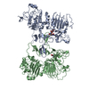
| Title | HER2 S310F in complex with TL1 Fab | ||||||
 Components Components |
| ||||||
 Keywords Keywords | SIGNALING PROTEIN / HER2 / Fab / Tyrosine kinase receptor | ||||||
| Function / homology |  Function and homology information Function and homology informationnegative regulation of immature T cell proliferation in thymus / ERBB3:ERBB2 complex / ERBB2-ERBB4 signaling pathway / GRB7 events in ERBB2 signaling / RNA polymerase I core binding / immature T cell proliferation in thymus / semaphorin receptor complex / ErbB-3 class receptor binding / motor neuron axon guidance / Sema4D induced cell migration and growth-cone collapse ...negative regulation of immature T cell proliferation in thymus / ERBB3:ERBB2 complex / ERBB2-ERBB4 signaling pathway / GRB7 events in ERBB2 signaling / RNA polymerase I core binding / immature T cell proliferation in thymus / semaphorin receptor complex / ErbB-3 class receptor binding / motor neuron axon guidance / Sema4D induced cell migration and growth-cone collapse / regulation of microtubule-based process / PLCG1 events in ERBB2 signaling / ERBB2-EGFR signaling pathway / neurotransmitter receptor localization to postsynaptic specialization membrane / enzyme-linked receptor protein signaling pathway / ERBB2 Activates PTK6 Signaling / neuromuscular junction development / ERBB2-ERBB3 signaling pathway / Drug-mediated inhibition of ERBB2 signaling / Resistance of ERBB2 KD mutants to trastuzumab / Resistance of ERBB2 KD mutants to sapitinib / Resistance of ERBB2 KD mutants to tesevatinib / Resistance of ERBB2 KD mutants to neratinib / Resistance of ERBB2 KD mutants to osimertinib / Resistance of ERBB2 KD mutants to afatinib / Resistance of ERBB2 KD mutants to AEE788 / Resistance of ERBB2 KD mutants to lapatinib / Drug resistance in ERBB2 TMD/JMD mutants / positive regulation of Rho protein signal transduction / positive regulation of MAP kinase activity / positive regulation of transcription by RNA polymerase I / ERBB2 Regulates Cell Motility / semaphorin-plexin signaling pathway / oligodendrocyte differentiation / PI3K events in ERBB2 signaling / positive regulation of protein targeting to membrane / regulation of angiogenesis / regulation of ERK1 and ERK2 cascade / Schwann cell development / coreceptor activity / Signaling by ERBB2 / myelination / TFAP2 (AP-2) family regulates transcription of growth factors and their receptors / transmembrane receptor protein tyrosine kinase activity / positive regulation of cell adhesion / GRB2 events in ERBB2 signaling / SHC1 events in ERBB2 signaling / cell surface receptor protein tyrosine kinase signaling pathway / basal plasma membrane / peptidyl-tyrosine phosphorylation / Constitutive Signaling by Overexpressed ERBB2 / Downregulation of ERBB2:ERBB3 signaling / cellular response to epidermal growth factor stimulus / positive regulation of translation / positive regulation of epithelial cell proliferation / phosphatidylinositol 3-kinase/protein kinase B signal transduction / neuromuscular junction / wound healing / Signaling by ERBB2 TMD/JMD mutants / receptor protein-tyrosine kinase / Signaling by ERBB2 ECD mutants / Signaling by ERBB2 KD Mutants / receptor tyrosine kinase binding / cellular response to growth factor stimulus / epidermal growth factor receptor signaling pathway / ruffle membrane / Downregulation of ERBB2 signaling / neuron differentiation / Constitutive Signaling by Aberrant PI3K in Cancer / transmembrane signaling receptor activity / PIP3 activates AKT signaling / myelin sheath / heart development / presynaptic membrane / PI5P, PP2A and IER3 Regulate PI3K/AKT Signaling / RAF/MAP kinase cascade / positive regulation of cell growth / protein tyrosine kinase activity / basolateral plasma membrane / early endosome / cell surface receptor signaling pathway / protein phosphorylation / cell population proliferation / receptor complex / positive regulation of MAPK cascade / endosome membrane / intracellular signal transduction / apical plasma membrane / protein heterodimerization activity / signaling receptor binding / negative regulation of apoptotic process / perinuclear region of cytoplasm / signal transduction / nucleoplasm / ATP binding / identical protein binding / nucleus / membrane / plasma membrane / cytosol Similarity search - Function | ||||||
| Biological species |  Homo sapiens (human) Homo sapiens (human) | ||||||
| Method | ELECTRON MICROSCOPY / single particle reconstruction / cryo EM / Resolution: 2.61 Å | ||||||
 Authors Authors | Bang, I. / Koide, S. | ||||||
| Funding support | 1items
| ||||||
 Citation Citation |  Journal: Nat Chem Biol / Year: 2025 Journal: Nat Chem Biol / Year: 2025Title: Selective targeting of oncogenic hotspot mutations of the HER2 extracellular domain. Authors: Injin Bang / Takamitsu Hattori / Nadia Leloup / Alexis Corrado / Atekana Nyamaa / Akiko Koide / Ken Geles / Elizabeth Buck / Shohei Koide /  Abstract: Oncogenic mutations in the extracellular domain (ECD) of cell-surface receptors could serve as tumor-specific antigens that are accessible to antibody therapeutics. Such mutations have been ...Oncogenic mutations in the extracellular domain (ECD) of cell-surface receptors could serve as tumor-specific antigens that are accessible to antibody therapeutics. Such mutations have been identified in receptor tyrosine kinases including HER2. However, it is challenging to selectively target a point mutant, while sparing the wild-type protein. Here we developed antibodies selective to HER2 S310F and S310Y, the two most common oncogenic mutations in the HER2 ECD, via combinatorial library screening and structure-guided design. Cryogenic-electron microscopy structures of the HER2 S310F homodimer and an antibody bound to HER2 S310F revealed that these antibodies recognize the mutations in a manner that mimics the dimerization arm of HER2 and thus inhibit HER2 dimerization. These antibodies as T cell engagers selectively killed a HER2 S310F-driven cancer cell line in vitro, and in vivo as a xenograft. These results validate HER2 ECD mutations as actionable therapeutic targets and offer promising candidates toward clinical development. | ||||||
| History |
|
- Structure visualization
| Structure viewer | Molecule:  Molmil Molmil Jmol/JSmol Jmol/JSmol |
|---|
- Downloads & links
- Download
| PDBx/mmCIF format |  8vqd.cif.gz 8vqd.cif.gz | 156.5 KB | Display |  PDBx/mmCIF format PDBx/mmCIF format |
|---|---|---|---|---|
| PDB format |  pdb8vqd.ent.gz pdb8vqd.ent.gz | 109.9 KB | Display |  PDB format PDB format |
| PDBx/mmJSON format |  8vqd.json.gz 8vqd.json.gz | Tree view |  PDBx/mmJSON format PDBx/mmJSON format | |
| Others |  Other downloads Other downloads |
-Validation report
| Arichive directory |  https://data.pdbj.org/pub/pdb/validation_reports/vq/8vqd https://data.pdbj.org/pub/pdb/validation_reports/vq/8vqd ftp://data.pdbj.org/pub/pdb/validation_reports/vq/8vqd ftp://data.pdbj.org/pub/pdb/validation_reports/vq/8vqd | HTTPS FTP |
|---|
-Related structure data
| Related structure data |  43439MC  8vqeC C: citing same article ( M: map data used to model this data |
|---|---|
| Similar structure data | Similarity search - Function & homology  F&H Search F&H Search |
- Links
Links
- Assembly
| Deposited unit | 
|
|---|---|
| 1 |
|
- Components
| #1: Protein | Mass: 99261.641 Da / Num. of mol.: 1 / Mutation: S310F Source method: isolated from a genetically manipulated source Source: (gene. exp.)  Homo sapiens (human) / Gene: ERBB2, HER2, MLN19, NEU, NGL / Production host: Homo sapiens (human) / Gene: ERBB2, HER2, MLN19, NEU, NGL / Production host:  Homo sapiens (human) Homo sapiens (human)References: UniProt: P04626, receptor protein-tyrosine kinase |
|---|---|
| #2: Antibody | Mass: 27512.574 Da / Num. of mol.: 1 Source method: isolated from a genetically manipulated source Source: (gene. exp.)  Homo sapiens (human) / Production host: Homo sapiens (human) / Production host:  |
| #3: Antibody | Mass: 23339.900 Da / Num. of mol.: 1 Source method: isolated from a genetically manipulated source Source: (gene. exp.)  Homo sapiens (human) / Production host: Homo sapiens (human) / Production host:  |
| #4: Polysaccharide | 2-acetamido-2-deoxy-beta-D-glucopyranose-(1-4)-2-acetamido-2-deoxy-beta-D-glucopyranose Source method: isolated from a genetically manipulated source |
| Has ligand of interest | N |
| Has protein modification | Y |
-Experimental details
-Experiment
| Experiment | Method: ELECTRON MICROSCOPY |
|---|---|
| EM experiment | Aggregation state: PARTICLE / 3D reconstruction method: single particle reconstruction |
- Sample preparation
| Component | Name: HER2 S310F in complex with TL1 Fab / Type: COMPLEX Details: Fab that only binds to HER2 S310F/Y but not to WT is in complex with HER2 S310F Entity ID: #1-#3 / Source: RECOMBINANT | ||||||||||||||||||||
|---|---|---|---|---|---|---|---|---|---|---|---|---|---|---|---|---|---|---|---|---|---|
| Molecular weight | Value: 150 kDa/nm / Experimental value: NO | ||||||||||||||||||||
| Source (natural) | Organism:  Homo sapiens (human) Homo sapiens (human) | ||||||||||||||||||||
| Source (recombinant) | Organism:  Homo sapiens (human) Homo sapiens (human) | ||||||||||||||||||||
| Buffer solution | pH: 7.5 Details: 20mM Tris, 150mM NaCl, 0.7mM fluorinated octyl maltoside | ||||||||||||||||||||
| Buffer component |
| ||||||||||||||||||||
| Specimen | Conc.: 1.4 mg/ml / Embedding applied: NO / Shadowing applied: NO / Staining applied: NO / Vitrification applied: YES Details: This sample was was eluted as a monodisperse peak with gel filtration | ||||||||||||||||||||
| Specimen support | Details: The grid was coated with goldfoil prior and discharged with current at 15mA for 25s, held for 10s. Grid material: COPPER / Grid mesh size: 300 divisions/in. / Grid type: Quantifoil R0.6/1 | ||||||||||||||||||||
| Vitrification | Instrument: FEI VITROBOT MARK IV / Cryogen name: ETHANE / Humidity: 95 % / Chamber temperature: 277.15 K Details: Vitrification carried out with blot force 5, blot time 4 s |
- Electron microscopy imaging
| Experimental equipment |  Model: Titan Krios / Image courtesy: FEI Company |
|---|---|
| Microscopy | Model: FEI TITAN KRIOS |
| Electron gun | Electron source:  FIELD EMISSION GUN / Accelerating voltage: 300 kV / Illumination mode: FLOOD BEAM FIELD EMISSION GUN / Accelerating voltage: 300 kV / Illumination mode: FLOOD BEAM |
| Electron lens | Mode: BRIGHT FIELD / Nominal magnification: 105000 X / Nominal defocus max: 2400 nm / Nominal defocus min: 900 nm / Cs: 2.7 mm |
| Image recording | Average exposure time: 2 sec. / Electron dose: 57.43 e/Å2 / Film or detector model: GATAN K3 BIOQUANTUM (6k x 4k) / Num. of grids imaged: 1 / Num. of real images: 8985 |
| EM imaging optics | Energyfilter name: GIF Bioquantum / Energyfilter slit width: 20 eV |
- Processing
| EM software |
| ||||||||||||||||||||||||||||||||||||||||||||||||||
|---|---|---|---|---|---|---|---|---|---|---|---|---|---|---|---|---|---|---|---|---|---|---|---|---|---|---|---|---|---|---|---|---|---|---|---|---|---|---|---|---|---|---|---|---|---|---|---|---|---|---|---|
| CTF correction | Type: PHASE FLIPPING AND AMPLITUDE CORRECTION | ||||||||||||||||||||||||||||||||||||||||||||||||||
| 3D reconstruction | Resolution: 2.61 Å / Resolution method: FSC 0.143 CUT-OFF / Num. of particles: 161060 / Symmetry type: POINT | ||||||||||||||||||||||||||||||||||||||||||||||||||
| Atomic model building | Protocol: FLEXIBLE FIT / Space: REAL | ||||||||||||||||||||||||||||||||||||||||||||||||||
| Atomic model building | 3D fitting-ID: 1
| ||||||||||||||||||||||||||||||||||||||||||||||||||
| Refine LS restraints |
|
 Movie
Movie Controller


 PDBj
PDBj


