Movie
Movie Controller

[English] 日本語
 Yorodumi
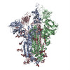
- PDB-8vpf: Structure of SARS-CoV spike in complex with CoV1-65 Fab (NTD-bound) -
+ Open data

- Basic information
| Entry | Database: PDB / ID: 8vpf | |||||||||||||||||||||||||||
|---|---|---|---|---|---|---|---|---|---|---|---|---|---|---|---|---|---|---|---|---|---|---|---|---|---|---|---|---|
| Title | Structure of SARS-CoV spike in complex with CoV1-65 Fab (NTD-bound) | |||||||||||||||||||||||||||
 Components Components |
| |||||||||||||||||||||||||||
 Keywords Keywords | VIRAL PROTEIN / SARS-CoV / Spike / Monoclonal antibody / Complex / NTD | |||||||||||||||||||||||||||
| Function / homology |  Function and homology information Function and homology informationMaturation of spike protein / Translation of Structural Proteins / Virion Assembly and Release / Attachment and Entry / SARS-CoV-1 activates/modulates innate immune responses / symbiont-mediated-mediated suppression of host tetherin activity / positive regulation of viral entry into host cell / membrane fusion / host cell endoplasmic reticulum-Golgi intermediate compartment membrane / receptor-mediated virion attachment to host cell ...Maturation of spike protein / Translation of Structural Proteins / Virion Assembly and Release / Attachment and Entry / SARS-CoV-1 activates/modulates innate immune responses / symbiont-mediated-mediated suppression of host tetherin activity / positive regulation of viral entry into host cell / membrane fusion / host cell endoplasmic reticulum-Golgi intermediate compartment membrane / receptor-mediated virion attachment to host cell / host cell surface receptor binding / symbiont-mediated suppression of host innate immune response / endocytosis involved in viral entry into host cell / fusion of virus membrane with host plasma membrane / fusion of virus membrane with host endosome membrane / viral envelope / host cell plasma membrane / virion membrane / membrane / identical protein binding Similarity search - Function | |||||||||||||||||||||||||||
| Biological species |  Severe acute respiratory syndrome coronavirus Severe acute respiratory syndrome coronavirus Homo sapiens (human) Homo sapiens (human) | |||||||||||||||||||||||||||
| Method | ELECTRON MICROSCOPY / single particle reconstruction / cryo EM / Resolution: 3.2 Å | |||||||||||||||||||||||||||
 Authors Authors | Bangaru, S. / Ward, A.B. | |||||||||||||||||||||||||||
| Funding support |  United States, 2items United States, 2items
| |||||||||||||||||||||||||||
 Citation Citation | Journal: J Clin Invest / Year: 2024 Title: Structural characterization of human monoclonal antibodies targeting uncommon antigenic sites on spike glycoprotein of SARS-CoV. Authors: Naveenchandra Suryadevara / Nurgun Kose / Sandhya Bangaru / Elad Binshtein / Jennifer Munt / David R Martinez / Alexandra Schäfer / Luke Myers / Trevor D Scobey / Robert H Carnahan / Andrew ...Authors: Naveenchandra Suryadevara / Nurgun Kose / Sandhya Bangaru / Elad Binshtein / Jennifer Munt / David R Martinez / Alexandra Schäfer / Luke Myers / Trevor D Scobey / Robert H Carnahan / Andrew B Ward / Ralph S Baric / James E Crowe / Abstract: The function of the spike protein N terminal domain (NTD) in coronavirus (CoV) infections is poorly understood. However, some rare antibodies that target the SARS-CoV-2 NTD potently neutralize the ...The function of the spike protein N terminal domain (NTD) in coronavirus (CoV) infections is poorly understood. However, some rare antibodies that target the SARS-CoV-2 NTD potently neutralize the virus. This finding suggests the NTD may contribute, in part, to protective immunity. Pansarbecovirus antibodies are desirable for broad protection, but the NTD region of SARS-CoV and SARS-CoV-2 exhibit a high level of sequence divergence; therefore, cross-reactive NTD-specific antibodies are unexpected, and there is no structure of a SARS-CoV NTD-specific antibody in complex with NTD. Here, we report a monoclonal antibody COV1-65, encoded by the IGHV1-69 gene, that recognizes the NTD of SARS-CoV S protein. A prophylaxis study showed the mAb COV1-65 prevented disease when administered before SARS-CoV challenge of BALB/c mice, an effect that requires intact fragment crystallizable region (Fc) effector functions for optimal protection in vivo. The footprint on the S protein of COV1-65 is near to functional components of the S2 fusion machinery, and the selection of COV1-65 escape mutant viruses identified critical residues Y886H and Q974H, which likely affect the epitope through allosteric effects. Structural features of the mAb COV1-65-SARS-CoV antigen interaction suggest critical antigenic determinants that should be considered in the rational design of sarbecovirus vaccine candidates. | |||||||||||||||||||||||||||
| History |
|
- Structure visualization
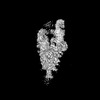
| Structure viewer | Molecule: MolmilJmol/JSmol |
|---|
- Downloads & links
-Download
| PDBx/mmCIF format | 8vpf.cif.gz | 623.7 KB | Display | PDBx/mmCIF format |
|---|---|---|---|---|
| PDB format | pdb8vpf.ent.gz | 489 KB | Display | PDB format |
| PDBx/mmJSON format | 8vpf.json.gz | Tree view | PDBx/mmJSON format | |
| Others |  Other downloads Other downloads |
-Validation report
| Arichive directory | https://data.pdbj.org/pub/pdb/validation_reports/vp/8vpfftp://data.pdbj.org/pub/pdb/validation_reports/vp/8vpf | HTTPS FTP |
|---|
-Related structure data
| Related structure data |  43407MC M: map data used to model this data C: citing same article ( |
|---|---|
| Similar structure data |
-Links
 PDBj
PDBj


- Assembly
| Deposited unit | 
|
|---|---|
| 1 |
|
-Components
-Protein , 1 types, 3 molecules ABC
| #1: Protein | Mass: 138740.688 Da / Num. of mol.: 3 Source method: isolated from a genetically manipulated source Source: (gene. exp.) Severe acute respiratory syndrome coronavirusGene: S, 2 / Cell line (production host): 293F / Production host: Homo sapiens (human) / References: UniProt: P59594 |
|---|
-Antibody , 2 types, 6 molecules MDENFG
| #2: Antibody | Mass: 24347.430 Da / Num. of mol.: 3 Source method: isolated from a genetically manipulated source Source: (gene. exp.) Homo sapiens (human) / Cell line (production host): 293F / Production host: Homo sapiens (human)#3: Antibody | Mass: 22878.279 Da / Num. of mol.: 3 Source method: isolated from a genetically manipulated source Source: (gene. exp.) Homo sapiens (human) / Cell line (production host): 293F / Production host: Homo sapiens (human) |
|---|
-Sugars , 3 types, 27 molecules 
| #4: Polysaccharide | 2-acetamido-2-deoxy-beta-D-glucopyranose-(1-4)-2-acetamido-2-deoxy-beta-D-glucopyranose Source method: isolated from a genetically manipulated source #5: Polysaccharide | Source method: isolated from a genetically manipulated source #6: Sugar | ChemComp-NAG /  Type: D-saccharide, beta linking / Mass: 221.208 Da / Num. of mol.: 15 / Source method: obtained synthetically / Formula: C8H15NO6 Type: D-saccharide, beta linking / Mass: 221.208 Da / Num. of mol.: 15 / Source method: obtained synthetically / Formula: C8H15NO6 |
|---|
-Details
| Has ligand of interest | N |
|---|---|
| Has protein modification | Y |
-Experimental details
-Experiment
| Experiment | Method: ELECTRON MICROSCOPY |
|---|---|
| EM experiment | Aggregation state: PARTICLE / 3D reconstruction method: single particle reconstruction |
- Sample preparation
| Component | Name: Structure of SARS-CoV spike in complex with CoV1-65 Fab Type: COMPLEX / Entity ID: #1-#3 / Source: MULTIPLE SOURCES | |||||||||||||||
|---|---|---|---|---|---|---|---|---|---|---|---|---|---|---|---|---|
| Molecular weight | Experimental value: NO | |||||||||||||||
| Source (natural) | Organism: Human coronavirus OC43 | |||||||||||||||
| Source (recombinant) | Organism: Homo sapiens (human) / Cell: 293F cells | |||||||||||||||
| Buffer solution | pH: 7.4 / Details: 1X TBS | |||||||||||||||
| Buffer component |
| |||||||||||||||
| Specimen | Conc.: 0.5 mg/ml / Embedding applied: NO / Shadowing applied: NO / Staining applied: NO / Vitrification applied: YES | |||||||||||||||
| Specimen support | Grid material: COPPER / Grid mesh size: 400 divisions/in. / Grid type: Quantifoil R2/1 | |||||||||||||||
| Vitrification | Instrument: FEI VITROBOT MARK IV / Cryogen name: ETHANE / Humidity: 100 % / Chamber temperature: 283 K |
- Electron microscopy imaging
| Experimental equipment |  Model: Titan Krios / Image courtesy: FEI Company |
|---|---|
| Microscopy | Model: FEI TITAN KRIOS |
| Electron gun | Electron source:  FIELD EMISSION GUN / Accelerating voltage: 300 kV / Illumination mode: FLOOD BEAM FIELD EMISSION GUN / Accelerating voltage: 300 kV / Illumination mode: FLOOD BEAM |
| Electron lens | Mode: BRIGHT FIELD / Nominal magnification: 29000 X / Nominal defocus max: 1600 nm / Nominal defocus min: 600 nm / Cs: 2.7 mm / C2 aperture diameter: 70 µm / Alignment procedure: COMA FREE |
| Specimen holder | Cryogen: NITROGEN / Specimen holder model: FEI TITAN KRIOS AUTOGRID HOLDER |
| Image recording | Electron dose: 50 e/Å2 / Detector mode: COUNTING / Film or detector model: GATAN K2 SUMMIT (4k x 4k) / Num. of real images: 2589 |
| Image scans | Movie frames/image: 30 |
- Processing
| EM software |
| |||||||||||||||||||||||||||||||||
|---|---|---|---|---|---|---|---|---|---|---|---|---|---|---|---|---|---|---|---|---|---|---|---|---|---|---|---|---|---|---|---|---|---|---|
| CTF correction | Type: PHASE FLIPPING AND AMPLITUDE CORRECTION | |||||||||||||||||||||||||||||||||
| Particle selection | Num. of particles selected: 315452 | |||||||||||||||||||||||||||||||||
| Symmetry | Point symmetry: C3 (3 fold cyclic) | |||||||||||||||||||||||||||||||||
| 3D reconstruction | Resolution: 3.2 Å / Resolution method: FSC 0.143 CUT-OFF / Num. of particles: 190300 / Symmetry type: POINT | |||||||||||||||||||||||||||||||||
| Atomic model building | PDB-ID: 6crx Accession code: 6crx / Source name: PDB / Type: experimental model |