 Movie
Movie Controller

[English] 日本語
 Yorodumi
- PDB-8vlg: Cryo-EM structure of human HGSNAT bound with Acetyl-CoA and subst... -
+ Open data

- Basic information
| Entry | Database: PDB / ID: 8vlg | |||||||||
|---|---|---|---|---|---|---|---|---|---|---|
| Title | Cryo-EM structure of human HGSNAT bound with Acetyl-CoA and substrate analog | |||||||||
 Components Components | Heparan-alpha-glucosaminide N-acetyltransferase | |||||||||
 Keywords Keywords | MEMBRANE PROTEIN / TRANSFERASE/SUBSTRATE / Lysosome / Transmembrane acetyltransferase / Acetyl-CoA / substrate / Heperan sulfate / Transferase-substrate analog complex / TRANSFERASE-SUBSTRATE complex | |||||||||
| Function / homology |  Function and homology information Function and homology informationheparan-alpha-glucosaminide N-acetyltransferase / heparan-alpha-glucosaminide N-acetyltransferase activity / MPS IIIC - Sanfilippo syndrome C / heparan sulfate proteoglycan catabolic process / HS-GAG degradation / lysosomal transport / acyltransferase activity / protein complex oligomerization / tertiary granule membrane / specific granule membrane ...heparan-alpha-glucosaminide N-acetyltransferase / heparan-alpha-glucosaminide N-acetyltransferase activity / MPS IIIC - Sanfilippo syndrome C / heparan sulfate proteoglycan catabolic process / HS-GAG degradation / lysosomal transport / acyltransferase activity / protein complex oligomerization / tertiary granule membrane / specific granule membrane / lysosomal lumen / lysosomal membrane / Neutrophil degranulation / plasma membrane Similarity search - Function | |||||||||
| Biological species |  Homo sapiens (human) Homo sapiens (human) | |||||||||
| Method | ELECTRON MICROSCOPY / single particle reconstruction / cryo EM / Resolution: 3.15 Å | |||||||||
 Authors Authors | Li, F. / Zhao, B. | |||||||||
| Funding support |  United States, 2items United States, 2items
| |||||||||
 Citation Citation | Journal: Nat Commun / Year: 2024 Title: Structural and mechanistic insights into a lysosomal membrane enzyme HGSNAT involved in Sanfilippo syndrome. Authors: Boyang Zhao / Zhongzheng Cao / Yi Zheng / Phuong Nguyen / Alisa Bowen / Robert H Edwards / Robert M Stroud / Yi Zhou / Menno Van Lookeren Campagne / Fei Li / Abstract: Heparan sulfate (HS) is degraded in lysosome by a series of glycosidases. Before the glycosidases can act, the terminal glucosamine of HS must be acetylated by the integral lysosomal membrane enzyme ...Heparan sulfate (HS) is degraded in lysosome by a series of glycosidases. Before the glycosidases can act, the terminal glucosamine of HS must be acetylated by the integral lysosomal membrane enzyme heparan-α-glucosaminide N-acetyltransferase (HGSNAT). Mutations of HGSNAT cause HS accumulation and consequently mucopolysaccharidosis IIIC, a devastating lysosomal storage disease characterized by progressive neurological deterioration and early death where no treatment is available. HGSNAT catalyzes a unique transmembrane acetylation reaction where the acetyl group of cytosolic acetyl-CoA is transported across the lysosomal membrane and attached to HS in one reaction. However, the reaction mechanism remains elusive. Here we report six cryo-EM structures of HGSNAT along the reaction pathway. These structures reveal a dimer arrangement and a unique structural fold, which enables the elucidation of the reaction mechanism. We find that a central pore within each monomer traverses the membrane and controls access of cytosolic acetyl-CoA to the active site at its luminal mouth where glucosamine binds. A histidine-aspartic acid catalytic dyad catalyzes the transfer reaction via a ternary complex mechanism. Furthermore, the structures allow the mapping of disease-causing variants and reveal their potential impact on the function, thus creating a framework to guide structure-based drug discovery efforts. | |||||||||
| History |
|
- Structure visualization
| Structure viewer | Molecule: MolmilJmol/JSmol |
|---|
- Downloads & links
-Download
| PDBx/mmCIF format | 8vlg.cif.gz | 222.6 KB | Display | PDBx/mmCIF format |
|---|---|---|---|---|
| PDB format | pdb8vlg.ent.gz | Display | PDB format | |
| PDBx/mmJSON format | 8vlg.json.gz | Tree view | PDBx/mmJSON format | |
| Others |  Other downloads Other downloads |
-Validation report
| Arichive directory | https://data.pdbj.org/pub/pdb/validation_reports/vl/8vlgftp://data.pdbj.org/pub/pdb/validation_reports/vl/8vlg | HTTPS FTP |
|---|
-Related structure data
| Related structure data |  43338MC  8vkjC  8vliC  8vluC 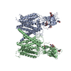 8vlvC  8vlyC M: map data used to model this data C: citing same article ( |
|---|---|
| Similar structure data |


-Links
 PDBj
PDBj- Assembly
| Deposited unit | 
|
|---|---|
| 1 |
|
-Components
| #1: Protein | Mass: 73365.039 Da / Num. of mol.: 2 Source method: isolated from a genetically manipulated source Source: (gene. exp.) Homo sapiens (human) / Gene: HGSNAT, TMEM76 / Production host: Homo sapiens (human)References: UniProt: Q68CP4, heparan-alpha-glucosaminide N-acetyltransferase #2: Polysaccharide | 2-acetamido-2-deoxy-beta-D-glucopyranose-(1-4)-2-acetamido-2-deoxy-beta-D-glucopyranose | Source method: isolated from a genetically manipulated source #3: Sugar | ChemComp-NAG /   Type: D-saccharide, beta linking / Mass: 221.208 Da / Num. of mol.: 4 Type: D-saccharide, beta linking / Mass: 221.208 Da / Num. of mol.: 4Source method: isolated from a genetically manipulated source Formula: C8H15NO6 #4: Chemical |   Mass: 809.571 Da / Num. of mol.: 2 / Source method: obtained synthetically / Formula: C23H38N7O17P3S / Feature type: SUBJECT OF INVESTIGATION Mass: 809.571 Da / Num. of mol.: 2 / Source method: obtained synthetically / Formula: C23H38N7O17P3S / Feature type: SUBJECT OF INVESTIGATION#5: Chemical | Mass: 337.325 Da / Num. of mol.: 2 / Source method: obtained synthetically / Formula: C16H19NO7 / Feature type: SUBJECT OF INVESTIGATION Has ligand of interest | Y | Has protein modification | Y | |
|---|
-Experimental details
-Experiment
| Experiment | Method: ELECTRON MICROSCOPY |
|---|---|
| EM experiment | Aggregation state: PARTICLE / 3D reconstruction method: single particle reconstruction |
- Sample preparation
| Component | Name: Purified human HGSNAT / Type: COMPLEX / Entity ID: #1 / Source: RECOMBINANT |
|---|---|
| Molecular weight | Value: 0.073 MDa / Experimental value: NO |
| Source (natural) | Organism: Homo sapiens (human) |
| Source (recombinant) | Organism: Homo sapiens (human) |
| Buffer solution | pH: 7.5 |
| Specimen | Conc.: 4.4 mg/ml / Embedding applied: NO / Shadowing applied: NO / Staining applied: NO / Vitrification applied: YES |
| Vitrification | Cryogen name: ETHANE |
- Electron microscopy imaging
| Experimental equipment |  Model: Titan Krios / Image courtesy: FEI Company |
|---|---|
| Microscopy | Model: FEI TITAN KRIOS |
| Electron gun | Electron source:  FIELD EMISSION GUN / Accelerating voltage: 300 kV / Illumination mode: FLOOD BEAM FIELD EMISSION GUN / Accelerating voltage: 300 kV / Illumination mode: FLOOD BEAM |
| Electron lens | Mode: BRIGHT FIELD / Nominal defocus max: 22000 nm / Nominal defocus min: 10000 nm |
| Image recording | Electron dose: 1 e/Å2 / Film or detector model: GATAN K3 BIOQUANTUM (6k x 4k) |
- Processing
| EM software | Name: PHENIX / Version: 1.20.1_4487: / Category: model refinement | ||||||||||||||||||||||||
|---|---|---|---|---|---|---|---|---|---|---|---|---|---|---|---|---|---|---|---|---|---|---|---|---|---|
| CTF correction | Type: PHASE FLIPPING AND AMPLITUDE CORRECTION | ||||||||||||||||||||||||
| 3D reconstruction | Resolution: 3.15 Å / Resolution method: FSC 0.143 CUT-OFF / Num. of particles: 202590 / Symmetry type: POINT | ||||||||||||||||||||||||
| Refine LS restraints |
|
