Movie
Movie Controller

[English] 日本語
 Yorodumi
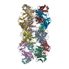
- PDB-8vgk: CryoEM structure of tryptase in complex with engineered conformat... -
+ Open data

- Basic information
| Entry | Database: PDB / ID: 8vgk | ||||||
|---|---|---|---|---|---|---|---|
| Title | CryoEM structure of tryptase in complex with engineered conformationally rigid anti-tryptase Fab E104.v1.6DS | ||||||
 Components Components |
| ||||||
 Keywords Keywords | HYDROLASE/IMMUNE SYSTEM / antibody fragment / fab / protein engineering / tryptase / HYDROLASE-IMMUNE SYSTEM complex | ||||||
| Function / homology |  Function and homology information Function and homology informationtryptase / Activation of Matrix Metalloproteinases / extracellular matrix disassembly / serine-type peptidase activity / defense response / serine-type endopeptidase activity / proteolysis / : / extracellular region / identical protein binding Similarity search - Function | ||||||
| Biological species |  Homo sapiens (human) Homo sapiens (human) | ||||||
| Method | ELECTRON MICROSCOPY / single particle reconstruction / cryo EM / Resolution: 2.4 Å | ||||||
 Authors Authors | Kung, J.E. / Johnson, M.C. / Sudhamsu, J. | ||||||
| Funding support | 1items
| ||||||
 Citation Citation | Journal: bioRxiv / Year: 2024 Title: Disulfi de constrained Fabs overcome target size limitation for high-resolution single-particle cryo-EM. Authors: Jennifer E Kung / Matthew C Johnson / Christine C Jao / Christopher P Arthur / Dimitry Tegunov / Alexis Rohou / Jawahar Sudhamsu /  Abstract: High-resolution structures of proteins are critical to understanding molecular mechanisms of biological processes and in the discovery of therapeutic molecules. Cryo-EM has revolutionized structure ...High-resolution structures of proteins are critical to understanding molecular mechanisms of biological processes and in the discovery of therapeutic molecules. Cryo-EM has revolutionized structure determination of large proteins and their complexes, but a vast majority of proteins that underlie human diseases are small (< 50 kDa) and usually beyond its reach due to low signal-to-noise images and difficulties in particle alignment. Current strategies to overcome this problem increase the overall size of small protein targets using scaffold proteins that bind to the target, but are limited by inherent flexibility and not being bound to their targets in a rigid manner, resulting in the target being poorly resolved compared to the scaffolds. Here we present an iteratively engineered molecular design for transforming Fabs (antibody fragments), into conformationally rigid scaffolds (Rigid-Fabs) that, when bound to small proteins (~20 kDa), can enable high-resolution structure determination using cryo-EM. This design introduces multiple disulfide bonds at strategic locations, generates a well-folded Fab constrained into a rigid conformation and can be applied to Fabs from various species, isotypes and chimeric Fabs. We present examples of the Rigid Fab design enabling high-resolution (2.3-2.5 Å) structures of small proteins, Ang2 (26 kDa) and KRAS (21 kDa) by cryo-EM. The strategies for designing disulfide constrained Rigid Fabs in our work thus establish a general approach to overcome the target size limitation of single particle cryo-EM. | ||||||
| History |
|
- Structure visualization
| Structure viewer | Molecule: MolmilJmol/JSmol |
|---|
- Downloads & links
-Download
| PDBx/mmCIF format | 8vgk.cif.gz | 504.5 KB | Display | PDBx/mmCIF format |
|---|---|---|---|---|
| PDB format | pdb8vgk.ent.gz | 413.6 KB | Display | PDB format |
| PDBx/mmJSON format | 8vgk.json.gz | Tree view | PDBx/mmJSON format | |
| Others |  Other downloads Other downloads |
-Validation report
| Arichive directory | https://data.pdbj.org/pub/pdb/validation_reports/vg/8vgkftp://data.pdbj.org/pub/pdb/validation_reports/vg/8vgk | HTTPS FTP |
|---|
-Related structure data
| Related structure data |  43203MC  8vegC  8vgeC  8vgfC  8vggC  8vghC 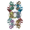 8vgiC 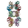 8vgjC 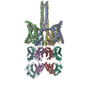 8vglC 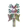 8vgmC 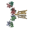 8vgnC 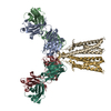 8vgoC 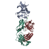 8vgpC 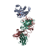 8vgqC M: map data used to model this data C: citing same article ( |
|---|---|
| Similar structure data |


-Links
 PDBj
PDBj


- Assembly
| Deposited unit | 
|
|---|---|
| 1 |
|
-Components
| #1: Protein | Mass: 27476.348 Da / Num. of mol.: 4 Source method: isolated from a genetically manipulated source Source: (gene. exp.) Homo sapiens (human) / Gene: TPSAB1, TPS1, TPS2, TPSB1 / Production host:  Trichoplusia ni (cabbage looper) / References: UniProt: Q15661, tryptase Trichoplusia ni (cabbage looper) / References: UniProt: Q15661, tryptase#2: Antibody | Mass: 23534.291 Da / Num. of mol.: 4 Source method: isolated from a genetically manipulated source Source: (gene. exp.) Homo sapiens (human) / Production host:   Cricetulus griseus (Chinese hamster) Cricetulus griseus (Chinese hamster)#3: Antibody | Mass: 25382.652 Da / Num. of mol.: 4 Source method: isolated from a genetically manipulated source Source: (gene. exp.) Homo sapiens (human) / Production host: Cricetulus griseus (Chinese hamster)#4: Water | ChemComp-HOH / |  Mass: 18.015 Da / Num. of mol.: 135 / Source method: isolated from a natural source / Formula: H2O Mass: 18.015 Da / Num. of mol.: 135 / Source method: isolated from a natural source / Formula: H2OHas protein modification | Y | |
|---|
-Experimental details
-Experiment
| Experiment | Method: ELECTRON MICROSCOPY |
|---|---|
| EM experiment | Aggregation state: PARTICLE / 3D reconstruction method: single particle reconstruction |
- Sample preparation
| Component |
| ||||||||||||||||||||||||
|---|---|---|---|---|---|---|---|---|---|---|---|---|---|---|---|---|---|---|---|---|---|---|---|---|---|
| Source (natural) |
| ||||||||||||||||||||||||
| Source (recombinant) |
| ||||||||||||||||||||||||
| Buffer solution | pH: 5.5 | ||||||||||||||||||||||||
| Buffer component |
| ||||||||||||||||||||||||
| Specimen | Embedding applied: NO / Shadowing applied: NO / Staining applied: NO / Vitrification applied: YES | ||||||||||||||||||||||||
| Specimen support | Details: Grid was treated overnight with 4 mM monothiolalkane(C11)PEG6-OH (11-mercaptoundecyl) hexaethyleneglycol then rinsed in ethanol prior to sample application. Grid material: GOLD / Grid mesh size: 300 divisions/in. / Grid type: Quantifoil R0.6/1 | ||||||||||||||||||||||||
| Vitrification | Instrument: FEI VITROBOT MARK IV / Cryogen name: ETHANE |
- Electron microscopy imaging
| Experimental equipment |  Model: Titan Krios / Image courtesy: FEI Company |
|---|---|
| Microscopy | Model: FEI TITAN KRIOS |
| Electron gun | Electron source:  FIELD EMISSION GUN / Accelerating voltage: 300 kV / Illumination mode: FLOOD BEAM FIELD EMISSION GUN / Accelerating voltage: 300 kV / Illumination mode: FLOOD BEAM |
| Electron lens | Mode: BRIGHT FIELD / Nominal magnification: 105000 X / Nominal defocus max: 1500 nm / Nominal defocus min: 500 nm / Cs: 2.7 mm / C2 aperture diameter: 70 µm |
| Image recording | Electron dose: 64 e/Å2 / Detector mode: COUNTING / Film or detector model: GATAN K3 BIOQUANTUM (6k x 4k) |
- Processing
| EM software |
| ||||||||||||||||||||||||||||||||||||||||
|---|---|---|---|---|---|---|---|---|---|---|---|---|---|---|---|---|---|---|---|---|---|---|---|---|---|---|---|---|---|---|---|---|---|---|---|---|---|---|---|---|---|
| CTF correction | Type: PHASE FLIPPING AND AMPLITUDE CORRECTION | ||||||||||||||||||||||||||||||||||||||||
| Symmetry | Point symmetry: D2 (2x2 fold dihedral) | ||||||||||||||||||||||||||||||||||||||||
| 3D reconstruction | Resolution: 2.4 Å / Resolution method: FSC 0.143 CUT-OFF / Num. of particles: 253508 / Symmetry type: POINT | ||||||||||||||||||||||||||||||||||||||||
| Atomic model building | Protocol: FLEXIBLE FIT / Space: REAL | ||||||||||||||||||||||||||||||||||||||||
| Atomic model building | PDB-ID: 6VVU Accession code: 6VVU / Source name: PDB / Type: experimental model | ||||||||||||||||||||||||||||||||||||||||
| Refine LS restraints |
|