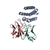
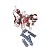
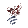
Journal: Nature / Year: 2024 Title: Broadly inhibitory antibodies to severe malaria virulence proteins. Authors: Raphael A Reyes / Sai Sundar Rajan Raghavan / Nicholas K Hurlburt / Viola Introini / Sebastiaan Bol / Ikhlaq Hussain Kana / Rasmus W Jensen / Elizabeth Martinez-Scholze / María Gestal-Mato ...Authors: Raphael A Reyes / Sai Sundar Rajan Raghavan / Nicholas K Hurlburt / Viola Introini / Sebastiaan Bol / Ikhlaq Hussain Kana / Rasmus W Jensen / Elizabeth Martinez-Scholze / María Gestal-Mato / Borja López-Gutiérrez / Silvia Sanz / Cristina Bancells / Monica Lisa Fernández-Quintero / Johannes R Loeffler / James Alexander Ferguson / Wen-Hsin Lee / Greg Michael Martin / Thor G Theander / John P A Lusingu / Daniel T R Minja / Isaac Ssewanyana / Margaret E Feeney / Bryan Greenhouse / Andrew B Ward / Maria Bernabeu / Marie Pancera / Louise Turner / Evelien M Bunnik / Thomas Lavstsen / Abstract: Malaria pathology is driven by the accumulation of Plasmodium falciparum-infected erythrocytes in microvessels. This process is mediated by the polymorphic erythrocyte membrane protein 1 (PfEMP1) ...Malaria pathology is driven by the accumulation of Plasmodium falciparum-infected erythrocytes in microvessels. This process is mediated by the polymorphic erythrocyte membrane protein 1 (PfEMP1) adhesion proteins of the parasite. A subset of PfEMP1 variants that bind to human endothelial protein C receptor (EPCR) through their CIDRα1 domains is responsible for severe malaria pathogenesis. A longstanding question is whether individual antibodies can recognize the large repertoire of circulating PfEMP1 variants. Here we describe two broadly reactive and inhibitory human monoclonal antibodies to CIDRα1. The antibodies isolated from two different individuals exhibited similar and consistent EPCR-binding inhibition of diverse CIDRα1 domains, representing five of the six subclasses of CIDRα1. Both antibodies inhibited EPCR binding of both recombinant full-length and native PfEMP1 proteins, as well as parasite sequestration in bioengineered 3D human brain microvessels under physiologically relevant flow conditions. Structural analyses of the two antibodies in complex with three different CIDRα1 antigen variants reveal similar binding mechanisms that depend on interactions with three highly conserved amino acid residues of the EPCR-binding site in CIDRα1. These broadly reactive antibodies are likely to represent a common mechanism of acquired immunity to severe malaria and offer novel insights for the design of a vaccine or treatment targeting severe malaria.
Mass: 19549.170 Da / Num. of mol.: 1 Source method: isolated from a genetically manipulated source Source: (gene. exp.) Plasmodium falciparum HB3 (eukaryote) / Cell line (production host): Expi293F / Production host: Homo sapiens (human)
#2: Antibody
C7HeavyChain
Mass: 25591.521 Da / Num. of mol.: 1 Source method: isolated from a genetically manipulated source Source: (gene. exp.) Homo sapiens (human) / Cell line (production host): HEK 293E / Production host: Homo sapiens (human)
#3: Protein
C7LightChain
Mass: 22939.492 Da / Num. of mol.: 1 Source method: isolated from a genetically manipulated source Source: (gene. exp.) Homo sapiens (human) / Cell line (production host): HEK 293E / Production host: Homo sapiens (human)
In the structure databanks used in Yorodumi, some data are registered as the other names, "COVID-19 virus" and "2019-nCoV". Here are the details of the virus and the list of structure data.
Jan 31, 2019. EMDB accession codes are about to change! (news from PDBe EMDB page)
EMDB accession codes are about to change! (news from PDBe EMDB page)
The allocation of 4 digits for EMDB accession codes will soon come to an end. Whilst these codes will remain in use, new EMDB accession codes will include an additional digit and will expand incrementally as the available range of codes is exhausted. The current 4-digit format prefixed with “EMD-” (i.e. EMD-XXXX) will advance to a 5-digit format (i.e. EMD-XXXXX), and so on. It is currently estimated that the 4-digit codes will be depleted around Spring 2019, at which point the 5-digit format will come into force.
The EM Navigator/Yorodumi systems omit the EMD- prefix.
Related info.:Q: What is EMD? / ID/Accession-code notation in Yorodumi/EM Navigator
Yorodumi is a browser for structure data from EMDB, PDB, SASBDB, etc.
This page is also the successor to EM Navigator detail page, and also detail information page/front-end page for Omokage search.
The word "yorodu" (or yorozu) is an old Japanese word meaning "ten thousand". "mi" (miru) is to see.
Related info.:EMDB / PDB / SASBDB / Comparison of 3 databanks / Yorodumi Search / Aug 31, 2016. New EM Navigator & Yorodumi / Yorodumi Papers / Jmol/JSmol / Function and homology information / Changes in new EM Navigator and Yorodumi
 Movie
Movie Controller


 Open data
 Basic information
 Components
 Keywords
 Homo sapiens (human)
 X-RAY DIFFRACTION /
  Authors
 United States, 1items
  Citation

 Structure visualization
 Molmil
 Downloads & links
 Other downloads
 PDBj
PDBj

 Assembly

 Mass: 65.409 Da / Num. of mol.: 6 / Source method: obtained synthetically / Formula: Zn
 Mass: 18.015 Da / Num. of mol.: 140 / Source method: isolated from a natural source / Formula: H2O
 Sample preparation
 Processing