 Movie
Movie Controller

[English] 日本語
 Yorodumi
- PDB-8vcq: Crystal structure of the oligomeric rMcL-1 in complex with raffinose -
+ Open data

- Basic information
| Entry | Database: PDB / ID: 8vcq | ||||||
|---|---|---|---|---|---|---|---|
| Title | Crystal structure of the oligomeric rMcL-1 in complex with raffinose | ||||||
 Components Components | Galactose-binding lectin | ||||||
 Keywords Keywords | SUGAR BINDING PROTEIN / Galactose binding lectin / rMcL-1 | ||||||
| Function / homology | galactose binding / raffinose / ACETATE ION / alpha-D-galactopyranose / Galactose-binding lectin Function and homology information Function and homology information | ||||||
| Biological species |  Mytilus californianus (California mussel) Mytilus californianus (California mussel) | ||||||
| Method |  X-RAY DIFFRACTION / MOLECULAR REPLACEMENT / Resolution: 2.09 Å X-RAY DIFFRACTION / MOLECULAR REPLACEMENT / Resolution: 2.09 Å | ||||||
 Authors Authors | Hernandez-Santoyo, A. / Loera-Rubalcava, J. | ||||||
| Funding support |  Mexico, 1items Mexico, 1items
| ||||||
 Citation Citation | Journal: Int.J.Biol.Macromol. / Year: 2025 Title: A mytilectin from Mytilus californianus: Study of its unique galactoside interactions, oligomerization patterns, and antifungal activity. Authors: Loera-Rubalcava, J. / Garcia-Maldonado, E. / Rodriguez-Romero, A. / Quintero-Martinez, A. / Macias-Rubalcava, M.L. / Cano-Sanchez, P. / Ramirez-Rodriguez, M.A. / Espinosa-Perez, G. / Hernandez-Santoyo, A. | ||||||
| History |
|
- Structure visualization
| Structure viewer | Molecule: MolmilJmol/JSmol |
|---|
- Downloads & links
-Download
| PDBx/mmCIF format | 8vcq.cif.gz | 299.6 KB | Display | PDBx/mmCIF format |
|---|---|---|---|---|
| PDB format | pdb8vcq.ent.gz | 227.2 KB | Display | PDB format |
| PDBx/mmJSON format | 8vcq.json.gz | Tree view | PDBx/mmJSON format | |
| Others |  Other downloads Other downloads |
-Validation report
| Arichive directory | https://data.pdbj.org/pub/pdb/validation_reports/vc/8vcqftp://data.pdbj.org/pub/pdb/validation_reports/vc/8vcq | HTTPS FTP |
|---|
-Related structure data
| Related structure data |  8vckC  8vcmC  8vcoC  8vcpC 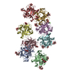 8vcsC  8vcuC C: citing same article ( |
|---|---|
| Similar structure data |
-Links
 PDBj
PDBj- Assembly
| Deposited unit | 
| ||||||||||||
|---|---|---|---|---|---|---|---|---|---|---|---|---|---|
| 1 |
| ||||||||||||
| Unit cell |
|
-Components
-Protein , 1 types, 8 molecules ABCDEFGH
| #1: Protein | Mass: 17225.705 Da / Num. of mol.: 8 Source method: isolated from a genetically manipulated source Source: (gene. exp.) Mytilus californianus (California mussel)Plasmid: pET-28a / Production host:  |
|---|
-Sugars , 2 types, 28 molecules 
| #2: Polysaccharide | alpha-D-galactopyranose-(1-6)-alpha-D-glucopyranose-(1-2)-beta-D-fructofuranose 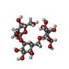  Source method: isolated from a genetically manipulated source Details: oligosaccharide with reducing-end-to-reducing-end glycosidic bond References: raffinose #3: Sugar |  Type: D-saccharide, alpha linking / Mass: 180.156 Da / Num. of mol.: 2 / Source method: obtained synthetically / Formula: C6H12O6 / Feature type: SUBJECT OF INVESTIGATION Type: D-saccharide, alpha linking / Mass: 180.156 Da / Num. of mol.: 2 / Source method: obtained synthetically / Formula: C6H12O6 / Feature type: SUBJECT OF INVESTIGATION |
|---|
-Non-polymers , 6 types, 363 molecules 


| #4: Chemical |  Mass: 238.305 Da / Num. of mol.: 2 / Source method: obtained synthetically / Formula: C8H18N2O4S / Comment: pH buffer*YM Mass: 238.305 Da / Num. of mol.: 2 / Source method: obtained synthetically / Formula: C8H18N2O4S / Comment: pH buffer*YM#5: Chemical |  Mass: 194.226 Da / Num. of mol.: 2 / Source method: obtained synthetically / Formula: C8H18O5 / Comment: precipitant*YM Mass: 194.226 Da / Num. of mol.: 2 / Source method: obtained synthetically / Formula: C8H18O5 / Comment: precipitant*YM#6: Chemical | ChemComp-CA / |  Mass: 40.078 Da / Num. of mol.: 1 / Source method: obtained synthetically / Formula: Ca Mass: 40.078 Da / Num. of mol.: 1 / Source method: obtained synthetically / Formula: Ca#7: Chemical | ChemComp-ACT / |  Mass: 59.044 Da / Num. of mol.: 1 / Source method: isolated from a natural source / Formula: C2H3O2 Mass: 59.044 Da / Num. of mol.: 1 / Source method: isolated from a natural source / Formula: C2H3O2#8: Chemical | ChemComp-GOL / |  Mass: 92.094 Da / Num. of mol.: 1 / Source method: obtained synthetically / Formula: C3H8O3 Mass: 92.094 Da / Num. of mol.: 1 / Source method: obtained synthetically / Formula: C3H8O3#9: Water | ChemComp-HOH / | Mass: 18.015 Da / Num. of mol.: 356 / Source method: isolated from a natural source / Formula: H2O |
|---|
-Details
| Has ligand of interest | Y |
|---|---|
| Has protein modification | N |
-Experimental details
-Experiment
| Experiment | Method: X-RAY DIFFRACTION / Number of used crystals: 1 |
|---|
- Sample preparation
| Crystal | Density Matthews: 2.81 Å3/Da / Density % sol: 56.18 % |
|---|---|
| Crystal grow | Temperature: 291.15 K / Method: vapor diffusion, sitting drop / pH: 7.5 Details: 18% PEG 4000, 50 mM sodium acetate, 0.1 M HEPES, 0.1 M lithium sulfate |
-Data collection
| Diffraction | Mean temperature: 101 K / Serial crystal experiment: N |
|---|---|
| Diffraction source | Source: ROTATING ANODE / Type: RIGAKU MICROMAX-007 HF / Wavelength: 1.54 Å |
| Detector | Type: DECTRIS PILATUS 200K / Detector: PIXEL / Date: Jan 20, 2017 |
| Radiation | Protocol: SINGLE WAVELENGTH / Monochromatic (M) / Laue (L): M / Scattering type: x-ray |
| Radiation wavelength | Wavelength: 1.54 Å / Relative weight: 1 |
| Reflection | Resolution: 2.09→49.34 Å / Num. obs: 159786 / % possible obs: 99.6 % / Redundancy: 7.8 % / Biso Wilson estimate: 36.97 Å2 / CC1/2: 0.804 / Rmerge(I) obs: 0.136 / Rpim(I) all: 0.048 / Rrim(I) all: 0.148 / Net I/σ(I): 29.2 |
| Reflection shell | Resolution: 2.09→2.11 Å / Redundancy: 5.5 % / Mean I/σ(I) obs: 2.3 / Num. unique obs: 3759 / CC1/2: 0.704 / % possible all: 97.5 |
- Processing
| Software |
| |||||||||||||||||||||||||||||||||||||||||||||||||||||||||||||||||||||||||||||||||||||||||||||||||||||||||||||||||||||||||||||||||||||||||||||||||||
|---|---|---|---|---|---|---|---|---|---|---|---|---|---|---|---|---|---|---|---|---|---|---|---|---|---|---|---|---|---|---|---|---|---|---|---|---|---|---|---|---|---|---|---|---|---|---|---|---|---|---|---|---|---|---|---|---|---|---|---|---|---|---|---|---|---|---|---|---|---|---|---|---|---|---|---|---|---|---|---|---|---|---|---|---|---|---|---|---|---|---|---|---|---|---|---|---|---|---|---|---|---|---|---|---|---|---|---|---|---|---|---|---|---|---|---|---|---|---|---|---|---|---|---|---|---|---|---|---|---|---|---|---|---|---|---|---|---|---|---|---|---|---|---|---|---|---|---|---|
| Refinement | Method to determine structure: MOLECULAR REPLACEMENT / Resolution: 2.09→49.34 Å / Cross valid method: FREE R-VALUE / σ(F): 1.34 / Phase error: 26.0499 Stereochemistry target values: GeoStd + Monomer Library + CDL v1.2
| |||||||||||||||||||||||||||||||||||||||||||||||||||||||||||||||||||||||||||||||||||||||||||||||||||||||||||||||||||||||||||||||||||||||||||||||||||
| Solvent computation | Shrinkage radii: 0.9 Å / VDW probe radii: 1.1 Å / Solvent model: FLAT BULK SOLVENT MODEL | |||||||||||||||||||||||||||||||||||||||||||||||||||||||||||||||||||||||||||||||||||||||||||||||||||||||||||||||||||||||||||||||||||||||||||||||||||
| Displacement parameters | Biso mean: 39.46 Å2 | |||||||||||||||||||||||||||||||||||||||||||||||||||||||||||||||||||||||||||||||||||||||||||||||||||||||||||||||||||||||||||||||||||||||||||||||||||
| Refinement step | Cycle: LAST / Resolution: 2.09→49.34 Å
| |||||||||||||||||||||||||||||||||||||||||||||||||||||||||||||||||||||||||||||||||||||||||||||||||||||||||||||||||||||||||||||||||||||||||||||||||||
| Refine LS restraints |
| |||||||||||||||||||||||||||||||||||||||||||||||||||||||||||||||||||||||||||||||||||||||||||||||||||||||||||||||||||||||||||||||||||||||||||||||||||
| LS refinement shell |
|
