 Movie
Movie Controller

[English] 日本語
 Yorodumi
- PDB-8va1: S. aureus TarL H300N in complex with CDP-ribitol (single tetramer) -
+ Open data

- Basic information
| Entry | Database: PDB / ID: 8va1 | |||||||||||||||
|---|---|---|---|---|---|---|---|---|---|---|---|---|---|---|---|---|
| Title | S. aureus TarL H300N in complex with CDP-ribitol (single tetramer) | |||||||||||||||
 Components Components | Teichoic acid ribitol-phosphate polymerase TarL | |||||||||||||||
 Keywords Keywords | TRANSFERASE / glycosyltransferase / polymerase / monotopic / amphipathic | |||||||||||||||
| Function / homology |  Function and homology information Function and homology informationCDP-ribitol ribitolphosphotransferase / CDP-ribitol ribitolphosphotransferase activity / CDP-glycerol glycerophosphotransferase activity / teichoic acid biosynthetic process / cell wall organization / plasma membrane Similarity search - Function | |||||||||||||||
| Biological species |   Staphylococcus aureus (bacteria) Staphylococcus aureus (bacteria) | |||||||||||||||
| Method | ELECTRON MICROSCOPY / single particle reconstruction / cryo EM / Resolution: 3.4 Å | |||||||||||||||
 Authors Authors | Li, F.K.K. / Worrall, L.J. / Strynadka, N.C.J. | |||||||||||||||
| Funding support |  Canada, Canada,  United States, 4items United States, 4items
| |||||||||||||||
 Citation Citation | Journal: Sci Adv / Year: 2024 Title: Cryo-EM analysis of TarL, a polymerase in wall teichoic acid biogenesis central to virulence and antibiotic resistance. Authors: Franco K K Li / Liam J Worrall / Robert T Gale / Eric D Brown / Natalie C J Strynadka / Abstract: Wall teichoic acid (WTA), a covalent adduct of Gram-positive bacterial cell wall peptidoglycan, contributes directly to virulence and antibiotic resistance in pathogenic species. Polymerization of ...Wall teichoic acid (WTA), a covalent adduct of Gram-positive bacterial cell wall peptidoglycan, contributes directly to virulence and antibiotic resistance in pathogenic species. Polymerization of the WTA ribitol-phosphate chain is catalyzed by TarL, a member of the largely uncharacterized TagF-like family of membrane-associated enzymes. We report the cryo-electron microscopy structure of TarL, showing a tetramer that forms an extensive membrane-binding platform of monotopic helices. TarL is composed of an amino-terminal immunoglobulin-like domain and a carboxyl-terminal glycosyltransferase-B domain for ribitol-phosphate polymerization. The active site of the latter is complexed to donor substrate cytidine diphosphate-ribitol, providing mechanistic insights into the catalyzed phosphotransfer reaction. Furthermore, the active site is surrounded by electropositive residues that serve to retain the lipid-linked acceptor for polymerization. Our data advance general insight into the architecture and membrane association of the still poorly characterized monotopic membrane protein class and present molecular details of ribitol-phosphate polymerization that may aid in the design of new antimicrobials. | |||||||||||||||
| History |
|
- Structure visualization
| Structure viewer | Molecule: MolmilJmol/JSmol |
|---|
- Downloads & links
-Download
| PDBx/mmCIF format | 8va1.cif.gz | 453.6 KB | Display | PDBx/mmCIF format |
|---|---|---|---|---|
| PDB format | pdb8va1.ent.gz | 375.4 KB | Display | PDB format |
| PDBx/mmJSON format | 8va1.json.gz | Tree view | PDBx/mmJSON format | |
| Others |  Other downloads Other downloads |
-Validation report
| Summary document | 8va1_validation.pdf.gz | 1.4 MB | Display | wwPDB validaton report |
|---|---|---|---|---|
| Full document | 8va1_full_validation.pdf.gz | 1.5 MB | Display | |
| Data in XML | 8va1_validation.xml.gz | 72.5 KB | Display | |
| Data in CIF | 8va1_validation.cif.gz | 104.2 KB | Display | |
| Arichive directory | https://data.pdbj.org/pub/pdb/validation_reports/va/8va1ftp://data.pdbj.org/pub/pdb/validation_reports/va/8va1 | HTTPS FTP |
-Related structure data
| Related structure data |  43083MC  8v33C 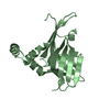 8v34C C: citing same article ( M: map data used to model this data |
|---|---|
| Similar structure data |

-Links
 PDBj
PDBj- Assembly
| Deposited unit | 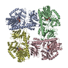
|
|---|---|
| 1 |
|
-Components
| #1: Protein | Mass: 68510.703 Da / Num. of mol.: 4 / Mutation: H300N Source method: isolated from a genetically manipulated source Source: (gene. exp.) Staphylococcus aureus (bacteria) / Gene: tarL, SAOUHSC_00227 / Production host: References: UniProt: Q2G1B8, CDP-ribitol ribitolphosphotransferase #2: Chemical | ChemComp-V2V / 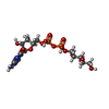  Mass: 537.307 Da / Num. of mol.: 4 / Source method: obtained synthetically / Formula: C14H25N3O15P2 / Feature type: SUBJECT OF INVESTIGATION Mass: 537.307 Da / Num. of mol.: 4 / Source method: obtained synthetically / Formula: C14H25N3O15P2 / Feature type: SUBJECT OF INVESTIGATIONHas ligand of interest | Y | |
|---|
-Experimental details
-Experiment
| Experiment | Method: ELECTRON MICROSCOPY |
|---|---|
| EM experiment | Aggregation state: PARTICLE / 3D reconstruction method: single particle reconstruction |
- Sample preparation
| Component | Name: S. aureus TarL H300N with CDP-ribitol and CHAPS / Type: COMPLEX / Entity ID: #1 / Source: RECOMBINANT | ||||||||||||||||
|---|---|---|---|---|---|---|---|---|---|---|---|---|---|---|---|---|---|
| Source (natural) | Organism: Staphylococcus aureus (bacteria) | ||||||||||||||||
| Source (recombinant) | Organism: | ||||||||||||||||
| Buffer solution | pH: 7.5 | ||||||||||||||||
| Buffer component |
| ||||||||||||||||
| Specimen | Conc.: 9.3 mg/ml / Embedding applied: NO / Shadowing applied: NO / Staining applied: NO / Vitrification applied: YES | ||||||||||||||||
| Specimen support | Grid material: COPPER / Grid mesh size: 300 divisions/in. | ||||||||||||||||
| Vitrification | Instrument: FEI VITROBOT MARK IV / Cryogen name: ETHANE / Humidity: 100 % / Chamber temperature: 277 K |
- Electron microscopy imaging
| Experimental equipment |  Model: Titan Krios / Image courtesy: FEI Company |
|---|---|
| Microscopy | Model: FEI TITAN KRIOS |
| Electron gun | Electron source:  FIELD EMISSION GUN / Accelerating voltage: 300 kV / Illumination mode: FLOOD BEAM FIELD EMISSION GUN / Accelerating voltage: 300 kV / Illumination mode: FLOOD BEAM |
| Electron lens | Mode: BRIGHT FIELD / Calibrated magnification: 81000 X / Nominal defocus max: 2500 nm / Nominal defocus min: 1000 nm |
| Image recording | Electron dose: 50 e/Å2 / Film or detector model: GATAN K3 (6k x 4k) / Num. of real images: 19380 |
- Processing
| EM software |
| ||||||||||||||||||||||||||||||||
|---|---|---|---|---|---|---|---|---|---|---|---|---|---|---|---|---|---|---|---|---|---|---|---|---|---|---|---|---|---|---|---|---|---|
| CTF correction | Type: PHASE FLIPPING AND AMPLITUDE CORRECTION | ||||||||||||||||||||||||||||||||
| Symmetry | Point symmetry: C1 (asymmetric) | ||||||||||||||||||||||||||||||||
| 3D reconstruction | Resolution: 3.4 Å / Resolution method: FSC 0.143 CUT-OFF / Num. of particles: 190753 / Symmetry type: POINT | ||||||||||||||||||||||||||||||||
| Atomic model building | PDB-ID: 3L7L Accession code: 3L7L / Source name: PDB / Type: experimental model | ||||||||||||||||||||||||||||||||
| Refinement | Cross valid method: NONE Stereochemistry target values: GeoStd + Monomer Library + CDL v1.2 | ||||||||||||||||||||||||||||||||
| Displacement parameters | Biso mean: 74.57 Å2 | ||||||||||||||||||||||||||||||||
| Refine LS restraints |
|

