 Movie
Movie Controller


+ Open data

- Basic information
| Entry | Database: PDB / ID: 8v6r | ||||||
|---|---|---|---|---|---|---|---|
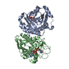
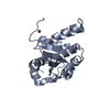
| Title | Crystal structure of EcThsA in complex with ADPR | ||||||
 Components Components | Thoeris protein ThsA Macro domain-containing protein | ||||||
 Keywords Keywords | IMMUNE SYSTEM / Antiphage immunity / nucleotide signalling | ||||||
| Function / homology | Thoeris protein ThsA, Macro domain / Thoeris protein ThsA, Macro domain / Chem-AR6 / Thoeris protein ThsA Macro domain-containing protein Function and homology information Function and homology information | ||||||
| Biological species |  | ||||||
| Method |  X-RAY DIFFRACTION / SYNCHROTRON / MOLECULAR REPLACEMENT / Resolution: 2.14 Å X-RAY DIFFRACTION / SYNCHROTRON / MOLECULAR REPLACEMENT / Resolution: 2.14 Å | ||||||
 Authors Authors | Shi, Y. / Masic, V. / Mosaiab, T. / Goulart, C.C. / Hartley-Tassell, L. / Sorbello, M. / Vasquez, E. / Mishra, B.P. / Holt, S. / Gu, W. ...Shi, Y. / Masic, V. / Mosaiab, T. / Goulart, C.C. / Hartley-Tassell, L. / Sorbello, M. / Vasquez, E. / Mishra, B.P. / Holt, S. / Gu, W. / Kobe, B. / Ve, T. | ||||||
| Funding support |  Australia, 1items Australia, 1items
| ||||||
 Citation Citation | Journal: Sci Adv / Year: 2024 Title: Structural characterization of macro domain-containing Thoeris antiphage defense systems. Authors: Shi, Y. / Masic, V. / Mosaiab, T. / Rajaratman, P. / Hartley-Tassell, L. / Sorbello, M. / Goulart, C.C. / Vasquez, E. / Mishra, B.P. / Holt, S. / Gu, W. / Kobe, B. / Ve, T. | ||||||
| History |
|
- Structure visualization
| Structure viewer | Molecule: MolmilJmol/JSmol |
|---|
- Downloads & links
-Download
| PDBx/mmCIF format | 8v6r.cif.gz | 324 KB | Display | PDBx/mmCIF format |
|---|---|---|---|---|
| PDB format | pdb8v6r.ent.gz | 222.7 KB | Display | PDB format |
| PDBx/mmJSON format | 8v6r.json.gz | Tree view | PDBx/mmJSON format | |
| Others |  Other downloads Other downloads |
-Validation report
| Arichive directory | https://data.pdbj.org/pub/pdb/validation_reports/v6/8v6rftp://data.pdbj.org/pub/pdb/validation_reports/v6/8v6r | HTTPS FTP |
|---|
-Related structure data

-Links
 PDBj
PDBj
- Assembly
| Deposited unit | 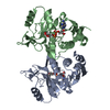
| ||||||||||||
|---|---|---|---|---|---|---|---|---|---|---|---|---|---|
| 1 |
| ||||||||||||
| Unit cell |
|
-Components
| #1: Protein | Mass: 27807.498 Da / Num. of mol.: 2 Source method: isolated from a genetically manipulated source Source: (gene. exp.) #2: Chemical |   Mass: 559.316 Da / Num. of mol.: 2 / Source method: obtained synthetically / Formula: C15H23N5O14P2 / Feature type: SUBJECT OF INVESTIGATION Mass: 559.316 Da / Num. of mol.: 2 / Source method: obtained synthetically / Formula: C15H23N5O14P2 / Feature type: SUBJECT OF INVESTIGATION#3: Water | ChemComp-HOH / |  Mass: 18.015 Da / Num. of mol.: 41 / Source method: isolated from a natural source / Formula: H2O Mass: 18.015 Da / Num. of mol.: 41 / Source method: isolated from a natural source / Formula: H2OHas ligand of interest | Y | |
|---|
-Experimental details
-Experiment
| Experiment | Method: X-RAY DIFFRACTION / Number of used crystals: 1 |
|---|
- Sample preparation
| Crystal | Density Matthews: 2.3 Å3/Da / Density % sol: 46.58 % |
|---|---|
| Crystal grow | Temperature: 293 K / Method: vapor diffusion, hanging drop Details: 14-24% w/v PEG 1000, 0.2 M lithium sulfate and 0.1 M phosphate-citrate buffer pH 3.8-4.2 |
-Data collection
| Diffraction | Mean temperature: 100 K / Serial crystal experiment: N |
|---|---|
| Diffraction source | Source: SYNCHROTRON / Site: Australian Synchrotron / Beamline: MX2 / Wavelength: 0.9537 Å |
| Detector | Type: DECTRIS EIGER X 16M / Detector: PIXEL / Date: Mar 20, 2020 |
| Radiation | Protocol: SINGLE WAVELENGTH / Monochromatic (M) / Laue (L): M / Scattering type: x-ray |
| Radiation wavelength | Wavelength: 0.9537 Å / Relative weight: 1 |
| Reflection | Resolution: 2.14→46.66 Å / Num. obs: 29133 / % possible obs: 99.8 % / Redundancy: 4.5 % / Biso Wilson estimate: 38.24 Å2 / CC1/2: 0.998 / Rmerge(I) obs: 0.099 / Rpim(I) all: 0.05 / Rrim(I) all: 0.111 / Net I/σ(I): 9 |
| Reflection shell | Resolution: 2.14→2.2 Å / Rmerge(I) obs: 0.969 / Num. unique obs: 2281 / CC1/2: 0.667 / Rpim(I) all: 0.49 / Rrim(I) all: 1.091 |
- Processing
| Software |
| |||||||||||||||||||||||||||||||||||||||||||||||||||||||||||||||||||||||||||||
|---|---|---|---|---|---|---|---|---|---|---|---|---|---|---|---|---|---|---|---|---|---|---|---|---|---|---|---|---|---|---|---|---|---|---|---|---|---|---|---|---|---|---|---|---|---|---|---|---|---|---|---|---|---|---|---|---|---|---|---|---|---|---|---|---|---|---|---|---|---|---|---|---|---|---|---|---|---|---|
| Refinement | Method to determine structure: MOLECULAR REPLACEMENT / Resolution: 2.14→41.67 Å / SU ML: 0.2356 / Cross valid method: FREE R-VALUE / σ(F): 1.34 / Phase error: 24.9167 Stereochemistry target values: GeoStd + Monomer Library + CDL v1.2
| |||||||||||||||||||||||||||||||||||||||||||||||||||||||||||||||||||||||||||||
| Solvent computation | Shrinkage radii: 0.9 Å / VDW probe radii: 1.11 Å / Solvent model: FLAT BULK SOLVENT MODEL | |||||||||||||||||||||||||||||||||||||||||||||||||||||||||||||||||||||||||||||
| Displacement parameters | Biso mean: 58.66 Å2 | |||||||||||||||||||||||||||||||||||||||||||||||||||||||||||||||||||||||||||||
| Refinement step | Cycle: LAST / Resolution: 2.14→41.67 Å
| |||||||||||||||||||||||||||||||||||||||||||||||||||||||||||||||||||||||||||||
| Refine LS restraints |
| |||||||||||||||||||||||||||||||||||||||||||||||||||||||||||||||||||||||||||||
| LS refinement shell |
| |||||||||||||||||||||||||||||||||||||||||||||||||||||||||||||||||||||||||||||
| Refinement TLS params. | Method: refined / Origin x: -15.0761647361 Å / Origin y: 19.3239526578 Å / Origin z: -18.4037750662 Å
| |||||||||||||||||||||||||||||||||||||||||||||||||||||||||||||||||||||||||||||
| Refinement TLS group | Selection details: all |
