 Movie
Movie Controller

[English] 日本語
 Yorodumi
- PDB-8v4g: X-ray structure of the NADP-dependent reductase from Campylobacte... -
+ Open data

- Basic information
| Entry | Database: PDB / ID: 8v4g | |||||||||
|---|---|---|---|---|---|---|---|---|---|---|
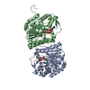
| Title | X-ray structure of the NADP-dependent reductase from Campylobacter jejuni responsible for the synthesis of CDP-glucitol in the presence of CDP and NADP | |||||||||
 Components Components | Putative nucleotide sugar dehydratase | |||||||||
 Keywords Keywords | OXIDOREDUCTASE / capsular polysaccharide / short chain dehydrogenase | |||||||||
| Function / homology | Oxidoreductases; Acting on the CH-OH group of donors; With NAD+ or NADP+ as acceptor / NAD-dependent epimerase/dehydratase / NAD dependent epimerase/dehydratase family / oxidoreductase activity / NAD(P)-binding domain superfamily / nucleotide binding / CYTIDINE-5'-DIPHOSPHATE / NADP NICOTINAMIDE-ADENINE-DINUCLEOTIDE PHOSPHATE / CDP-6-D-glucitol synthase Function and homology information Function and homology information | |||||||||
| Biological species |   Campylobacter jejuni (Campylobacter) Campylobacter jejuni (Campylobacter) | |||||||||
| Method |  X-RAY DIFFRACTION / MOLECULAR REPLACEMENT / Resolution: 2 Å X-RAY DIFFRACTION / MOLECULAR REPLACEMENT / Resolution: 2 Å | |||||||||
 Authors Authors | Schumann, M.E. / Thoden, J.B. / Holden, H.M. / Raushel, F.M. | |||||||||
| Funding support |  United States, 2items United States, 2items
| |||||||||
 Citation Citation | Journal: Biochemistry / Year: 2024 Title: Biosynthesis of Cytidine Diphosphate-6-d-Glucitol for the Capsular Polysaccharides of Campylobacter jejuni. Authors: Ghosh, M.K. / Narindoshvili, T. / Thoden, J.B. / Schumann, M.E. / Holden, H.M. / Raushel, F.M. | |||||||||
| History |
|
- Structure visualization
| Structure viewer | Molecule: MolmilJmol/JSmol |
|---|
- Downloads & links
-Download
| PDBx/mmCIF format | 8v4g.cif.gz | 155.1 KB | Display | PDBx/mmCIF format |
|---|---|---|---|---|
| PDB format | pdb8v4g.ent.gz | 119.8 KB | Display | PDB format |
| PDBx/mmJSON format | 8v4g.json.gz | Tree view | PDBx/mmJSON format | |
| Others |  Other downloads Other downloads |
-Validation report
| Summary document | 8v4g_validation.pdf.gz | 1.7 MB | Display | wwPDB validaton report |
|---|---|---|---|---|
| Full document | 8v4g_full_validation.pdf.gz | 1.7 MB | Display | |
| Data in XML | 8v4g_validation.xml.gz | 28.3 KB | Display | |
| Data in CIF | 8v4g_validation.cif.gz | 39.8 KB | Display | |
| Arichive directory | https://data.pdbj.org/pub/pdb/validation_reports/v4/8v4gftp://data.pdbj.org/pub/pdb/validation_reports/v4/8v4g | HTTPS FTP |
-Related structure data
-Links
 PDBj
PDBj- Assembly
| Deposited unit | 
| ||||||||
|---|---|---|---|---|---|---|---|---|---|
| 1 |
| ||||||||
| Unit cell |
|
-Components
-Protein , 1 types, 2 molecules AB
| #1: Protein | Mass: 38876.953 Da / Num. of mol.: 2 Source method: isolated from a genetically manipulated source Source: (gene. exp.) Campylobacter jejuni (Campylobacter) / Strain: HS:5 / Production host: |
|---|
-Non-polymers , 5 types, 245 molecules 
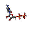


| #2: Chemical |  Mass: 743.405 Da / Num. of mol.: 2 / Source method: obtained synthetically / Formula: C21H28N7O17P3 / Feature type: SUBJECT OF INVESTIGATION Mass: 743.405 Da / Num. of mol.: 2 / Source method: obtained synthetically / Formula: C21H28N7O17P3 / Feature type: SUBJECT OF INVESTIGATION#3: Chemical |  Mass: 403.176 Da / Num. of mol.: 2 / Source method: obtained synthetically / Formula: C9H15N3O11P2 / Feature type: SUBJECT OF INVESTIGATION Mass: 403.176 Da / Num. of mol.: 2 / Source method: obtained synthetically / Formula: C9H15N3O11P2 / Feature type: SUBJECT OF INVESTIGATION#4: Chemical |  Mass: 35.453 Da / Num. of mol.: 2 / Source method: obtained synthetically / Formula: Cl Mass: 35.453 Da / Num. of mol.: 2 / Source method: obtained synthetically / Formula: Cl#5: Chemical |  Mass: 62.068 Da / Num. of mol.: 2 / Source method: obtained synthetically / Formula: C2H6O2 Mass: 62.068 Da / Num. of mol.: 2 / Source method: obtained synthetically / Formula: C2H6O2#6: Water | ChemComp-HOH / | Mass: 18.015 Da / Num. of mol.: 237 / Source method: isolated from a natural source / Formula: H2O |
|---|
-Details
| Has ligand of interest | Y |
|---|
-Experimental details
-Experiment
| Experiment | Method: X-RAY DIFFRACTION / Number of used crystals: 1 |
|---|
- Sample preparation
| Crystal | Density Matthews: 2.35 Å3/Da / Density % sol: 47.66 % |
|---|---|
| Crystal grow | Temperature: 293 K / Method: vapor diffusion, hanging drop / pH: 9 Details: protein incubated with 5 mM CDP and 5 mM NADP. Precipitant: 10-14 % w/v poly(ethylene glycol) 8000, 2% v/v hexyleneglycol, and 100 mM CHES (pH 9.0) |
-Data collection
| Diffraction | Mean temperature: 100 K / Serial crystal experiment: N |
|---|---|
| Diffraction source | Source: SEALED TUBE / Type: BRUKER D8 QUEST / Wavelength: 1.54178 Å |
| Detector | Type: Bruker PHOTON II / Detector: PIXEL / Date: Apr 5, 2023 |
| Radiation | Protocol: SINGLE WAVELENGTH / Monochromatic (M) / Laue (L): M / Scattering type: x-ray |
| Radiation wavelength | Wavelength: 1.54178 Å / Relative weight: 1 |
| Reflection | Resolution: 2→50 Å / Num. obs: 49653 / % possible obs: 98.4 % / Observed criterion σ(F): 0 / Observed criterion σ(I): 0 / Redundancy: 11.8 % / Rsym value: 0.094 / Net I/σ(I): 16.5 |
| Reflection shell | Resolution: 2→2.1 Å / Redundancy: 5.1 % / Mean I/σ(I) obs: 2.6 / Num. unique obs: 6294 / Rsym value: 0.46 / % possible all: 94.5 |
- Processing
| Software |
| ||||||||||||||||||||||||||||||||||||||||||||||||||||||||||||||||||||||||||||||||||||||||||||||||||||||||||||||||||||||||||||||||||||||||||||||||||||||||||||||||||||||||||||||||||||||
|---|---|---|---|---|---|---|---|---|---|---|---|---|---|---|---|---|---|---|---|---|---|---|---|---|---|---|---|---|---|---|---|---|---|---|---|---|---|---|---|---|---|---|---|---|---|---|---|---|---|---|---|---|---|---|---|---|---|---|---|---|---|---|---|---|---|---|---|---|---|---|---|---|---|---|---|---|---|---|---|---|---|---|---|---|---|---|---|---|---|---|---|---|---|---|---|---|---|---|---|---|---|---|---|---|---|---|---|---|---|---|---|---|---|---|---|---|---|---|---|---|---|---|---|---|---|---|---|---|---|---|---|---|---|---|---|---|---|---|---|---|---|---|---|---|---|---|---|---|---|---|---|---|---|---|---|---|---|---|---|---|---|---|---|---|---|---|---|---|---|---|---|---|---|---|---|---|---|---|---|---|---|---|---|
| Refinement | Method to determine structure: MOLECULAR REPLACEMENT / Resolution: 2→43.05 Å / Cor.coef. Fo:Fc: 0.948 / Cor.coef. Fo:Fc free: 0.907 / SU B: 5.187 / SU ML: 0.139 / Cross valid method: THROUGHOUT / ESU R: 0.191 / ESU R Free: 0.181 / Stereochemistry target values: MAXIMUM LIKELIHOOD / Details: HYDROGENS HAVE BEEN ADDED IN THE RIDING POSITIONS
| ||||||||||||||||||||||||||||||||||||||||||||||||||||||||||||||||||||||||||||||||||||||||||||||||||||||||||||||||||||||||||||||||||||||||||||||||||||||||||||||||||||||||||||||||||||||
| Solvent computation | Ion probe radii: 0.8 Å / Shrinkage radii: 0.8 Å / VDW probe radii: 1.2 Å / Solvent model: MASK | ||||||||||||||||||||||||||||||||||||||||||||||||||||||||||||||||||||||||||||||||||||||||||||||||||||||||||||||||||||||||||||||||||||||||||||||||||||||||||||||||||||||||||||||||||||||
| Displacement parameters | Biso mean: 26.348 Å2
| ||||||||||||||||||||||||||||||||||||||||||||||||||||||||||||||||||||||||||||||||||||||||||||||||||||||||||||||||||||||||||||||||||||||||||||||||||||||||||||||||||||||||||||||||||||||
| Refinement step | Cycle: 1 / Resolution: 2→43.05 Å
| ||||||||||||||||||||||||||||||||||||||||||||||||||||||||||||||||||||||||||||||||||||||||||||||||||||||||||||||||||||||||||||||||||||||||||||||||||||||||||||||||||||||||||||||||||||||
| Refine LS restraints |
|