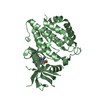
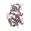
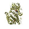
- PDB-8v42: Structure of Human Vaccinia-related Kinase 1 (VRK1) Bound to ACH000400 -
+
Open data
ID or keywords:
Loading...
-
Basic information
Entry
Database: PDB / ID: 8v42
Title
Structure of Human Vaccinia-related Kinase 1 (VRK1) Bound to ACH000400
Components
Serine/threonine-protein kinase VRK1
Keywords
TRANSFERASE / protein kinase / inhibitor / co-crystal
Function / homology
Function and homology information
histone H2AX kinase activity / Golgi disassembly / Cajal body organization / histone H3T3 kinase activity / Nuclear Envelope Breakdown / positive regulation of protein localization to chromatin / mitotic nuclear membrane disassembly / histone H3S10 kinase activity / regulation of neuron migration / Initiation of Nuclear Envelope (NE) Reformation ...histone H2AX kinase activity / Golgi disassembly / Cajal body organization / histone H3T3 kinase activity / Nuclear Envelope Breakdown / positive regulation of protein localization to chromatin / mitotic nuclear membrane disassembly / histone H3S10 kinase activity / regulation of neuron migration / Initiation of Nuclear Envelope (NE) Reformation / Golgi stack / nucleosomal DNA binding / Cajal body / neuron projection development / kinase activity / protein autophosphorylation / histone binding / protein phosphorylation / non-specific serine/threonine protein kinase / protein kinase activity / chromatin remodeling / cell division / protein serine kinase activity / protein serine/threonine kinase activity / DNA damage response / protein kinase binding / chromatin / nucleolus / signal transduction / nucleoplasm / ATP binding / nucleus / cytoplasm / cytosol Similarity search - Function
: / Serine/threonine-protein kinase, active site / Serine/Threonine protein kinases active-site signature. / Protein kinase domain / Serine/Threonine protein kinases, catalytic domain / Protein kinase, ATP binding site / Protein kinases ATP-binding region signature. / Protein kinase domain profile. / Protein kinase domain / Protein kinase-like domain superfamily Similarity search - Domain/homology
Monochromator: M / Protocol: SINGLE WAVELENGTH / Monochromatic (M) / Laue (L): M / Scattering type: x-ray
Radiation wavelength
Wavelength: 0.9686 Å / Relative weight: 1
Reflection
Resolution: 2.3→19.93 Å / Num. obs: 75682 / % possible obs: 99.7 % / Redundancy: 13.6 % / Rmerge(I) obs: 0.176 / Net I/σ(I): 11.8
Reflection shell
Resolution: 2.3→2.35 Å / Redundancy: 13.8 % / Rmerge(I) obs: 2.417 / Mean I/σ(I) obs: 1.8 / % possible all: 99.6
-
Processing
Software
Name
Version
Classification
REFMAC
5.8.0425
refinement
Aimless
datascaling
XDS
datareduction
PHASER
phasing
Refinement
Method to determine structure: MOLECULAR REPLACEMENT / Resolution: 2.3→19.93 Å / Cor.coef. Fo:Fc: 0.956 / Cor.coef. Fo:Fc free: 0.944 / SU B: 14.927 / SU ML: 0.166 / Cross valid method: THROUGHOUT / ESU R: 0.259 / ESU R Free: 0.201 Details: HYDROGENS HAVE BEEN ADDED IN THEIR RIDING POSITIONS
Rfactor
Num. reflection
% reflection
Selection details
Rfree
0.231
3800
5.027 %
RANDOM
Rwork
0.199
-
-
-
obs
-
75597
99.6 %
-
Solvent computation
Ion probe radii: 0.8 Å / Shrinkage radii: 0.8 Å / VDW probe radii: 1.2 Å / Solvent model: MASK BULK SOLVENT
 Movie
Movie Controller

 Yorodumi
 Open data
 Basic information
 Components
 Keywords
 Function and homology information
 Homo sapiens (human)
 X-RAY DIFFRACTION /
  Authors
 Brazil, 1items
  Citation
 Structure visualization
 Downloads & links
 Other downloads
 PDBj
PDBj

 Assembly
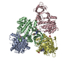


 Mass: 106.120 Da / Num. of mol.: 1 / Source method: obtained synthetically / Formula: C4H10O3
 Mass: 96.063 Da / Num. of mol.: 4 / Source method: obtained synthetically / Formula: SO4
 Mass: 18.015 Da / Num. of mol.: 142 / Source method: isolated from a natural source / Formula: H2O
 Sample preparation
 / Beamline: I24 / Wavelength: 0.9686 Å
 Processing