 Movie
Movie Controller


+ Open data

- Basic information
| Entry | Database: PDB / ID: 8uzw | ||||||||||||||||||
|---|---|---|---|---|---|---|---|---|---|---|---|---|---|---|---|---|---|---|---|
| Title | Selenocysteine synthase- SelA | ||||||||||||||||||
 Components Components | L-seryl-tRNA(Sec) selenium transferase | ||||||||||||||||||
 Keywords Keywords | RNA BINDING PROTEIN / Selenocysteine / tRNASec / Sec-synthase | ||||||||||||||||||
| Function / homology |  Function and homology information Function and homology informationL-seryl-tRNASec selenium transferase / L-seryl-tRNA(Sec) selenium transferase activity / conversion of seryl-tRNAsec to selenocys-tRNAsec / L-selenocysteine biosynthetic process / selenocysteine incorporation / pyridoxal phosphate binding / identical protein binding / cytosol Similarity search - Function | ||||||||||||||||||
| Biological species |  | ||||||||||||||||||
| Method | ELECTRON MICROSCOPY / single particle reconstruction / cryo EM / Resolution: 2.69 Å | ||||||||||||||||||
 Authors Authors | Balasco Serrao, V.H. / Minari, K. / Pereira, H.M. / Thiemann, O.H. | ||||||||||||||||||
| Funding support |  Brazil, Brazil,  United States, 5items United States, 5items
| ||||||||||||||||||
 Citation Citation | Journal: Curr Res Struct Biol / Year: 2024 Title: Bacterial selenocysteine synthase structure revealed by single-particle cryoEM. Authors: Vitor Hugo Balasco Serrão / Karine Minari / Humberto D'Muniz Pereira / Otavio Henrique Thiemann / Abstract: The 21st amino acid, selenocysteine (Sec), is synthesized on its dedicated transfer RNA (tRNA). In bacteria, Sec is synthesized from Ser-tRNA by Selenocysteine Synthase (SelA), which is a pivotal ...The 21st amino acid, selenocysteine (Sec), is synthesized on its dedicated transfer RNA (tRNA). In bacteria, Sec is synthesized from Ser-tRNA by Selenocysteine Synthase (SelA), which is a pivotal enzyme in the biosynthesis of Sec. The structural characterization of bacterial SelA is of paramount importance to decipher its catalytic mechanism and its role in the regulation of the Sec-synthesis pathway. Here, we present a comprehensive single-particle cryo-electron microscopy (SPA cryoEM) structure of the bacterial SelA with an overall resolution of 2.69 Å. Using recombinant SelA, we purified and prepared samples for single-particle cryoEM. The structural insights from SelA, combined with previous and knowledge, underscore the indispensable role of decamerization in SelA's function. Moreover, our structural analysis corroborates previous results that show that SelA adopts a pentamer of dimers configuration, and the active site architecture, substrate binding pocket, and key K295 catalytic residue are identified and described in detail. The differences in protein architecture and substrate coordination between the bacterial enzyme and its counterparts offer compelling structural evidence supporting the independent molecular evolution of the bacterial and archaea/eukarya Ser-Sec biosynthesis present in the natural world. | ||||||||||||||||||
| History |
|
- Structure visualization
| Structure viewer | Molecule: MolmilJmol/JSmol |
|---|
- Downloads & links
-Download
| PDBx/mmCIF format | 8uzw.cif.gz | 689.4 KB | Display | PDBx/mmCIF format |
|---|---|---|---|---|
| PDB format | pdb8uzw.ent.gz | 570 KB | Display | PDB format |
| PDBx/mmJSON format | 8uzw.json.gz | Tree view | PDBx/mmJSON format | |
| Others |  Other downloads Other downloads |
-Validation report
| Arichive directory | https://data.pdbj.org/pub/pdb/validation_reports/uz/8uzwftp://data.pdbj.org/pub/pdb/validation_reports/uz/8uzw | HTTPS FTP |
|---|
-Related structure data
| Related structure data |  42845MC M: map data used to model this data C: citing same article ( |
|---|---|
| Similar structure data | |
| Experimental dataset #1 | Data reference: 10.6019/EMPIAR-12043 / Data set type: EMPIAR |
-Links
 PDBj
PDBj- Assembly
| Deposited unit | 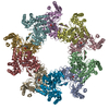
|
|---|---|
| 1 |
|
-Components
| #1: Protein | Mass: 50895.172 Da / Num. of mol.: 10 Source method: isolated from a genetically manipulated source Source: (gene. exp.) References: UniProt: P0A821, L-seryl-tRNASec selenium transferase Has ligand of interest | N | |
|---|
-Experimental details
-Experiment
| Experiment | Method: ELECTRON MICROSCOPY |
|---|---|
| EM experiment | Aggregation state: PARTICLE / 3D reconstruction method: single particle reconstruction |
- Sample preparation
| Component | Name: Bacterial Selenocysteine Synthase / Type: COMPLEX Details: Heterologously expressed E. coli homodecameric Selenocysteine Synthase Entity ID: all / Source: RECOMBINANT | |||||||||||||||||||||||||
|---|---|---|---|---|---|---|---|---|---|---|---|---|---|---|---|---|---|---|---|---|---|---|---|---|---|---|
| Molecular weight | Value: 0.51 MDa / Experimental value: NO | |||||||||||||||||||||||||
| Source (natural) | Organism: | |||||||||||||||||||||||||
| Source (recombinant) | Organism: | |||||||||||||||||||||||||
| Buffer solution | pH: 7.5 Details: 20 mM potassium phosphate pH 7.5, 100 mM sodium chloride, 2 mM 2-mercaptoethanol, and 10 uM pyridoxal 5-phosphate. | |||||||||||||||||||||||||
| Buffer component |
| |||||||||||||||||||||||||
| Specimen | Conc.: 3.9 mg/ml / Embedding applied: NO / Shadowing applied: NO / Staining applied: NO / Vitrification applied: YES Details: Monodisperse sample after size-exclusion chromatography | |||||||||||||||||||||||||
| Specimen support | Grid material: COPPER / Grid mesh size: 400 divisions/in. / Grid type: Quantifoil | |||||||||||||||||||||||||
| Vitrification | Instrument: FEI VITROBOT MARK IV / Cryogen name: ETHANE / Humidity: 100 % / Chamber temperature: 295 K Details: The sample was blotted for 2.5 seconds using a force of -10 and then swiftly plunged frozen into liquid ethane using the Vitrobot Mark IV - Thermo Fisher Scientific. |
- Electron microscopy imaging
| Experimental equipment |  Model: Titan Krios / Image courtesy: FEI Company |
|---|---|
| Microscopy | Model: FEI TITAN KRIOS |
| Electron gun | Electron source:  FIELD EMISSION GUN / Accelerating voltage: 300 kV / Illumination mode: FLOOD BEAM FIELD EMISSION GUN / Accelerating voltage: 300 kV / Illumination mode: FLOOD BEAM |
| Electron lens | Mode: BRIGHT FIELD / Nominal defocus max: 2800 nm / Nominal defocus min: 600 nm |
| Image recording | Electron dose: 45.4 e/Å2 / Film or detector model: FEI FALCON IV (4k x 4k) / Num. of grids imaged: 1 / Num. of real images: 4592 |
- Processing
| EM software |
| ||||||||||||||||||||||||||||||||||||||||
|---|---|---|---|---|---|---|---|---|---|---|---|---|---|---|---|---|---|---|---|---|---|---|---|---|---|---|---|---|---|---|---|---|---|---|---|---|---|---|---|---|---|
| CTF correction | Type: PHASE FLIPPING AND AMPLITUDE CORRECTION | ||||||||||||||||||||||||||||||||||||||||
| Particle selection | Num. of particles selected: 688602 | ||||||||||||||||||||||||||||||||||||||||
| Symmetry | Point symmetry: D5 (2x5 fold dihedral) | ||||||||||||||||||||||||||||||||||||||||
| 3D reconstruction | Resolution: 2.69 Å / Resolution method: FSC 0.143 CUT-OFF / Num. of particles: 223410 / Symmetry type: POINT | ||||||||||||||||||||||||||||||||||||||||
| Atomic model building | B value: 95.2 / Protocol: RIGID BODY FIT / Space: REAL | ||||||||||||||||||||||||||||||||||||||||
| Refine LS restraints |
|
