 Movie
Movie Controller


+ Open data

- Basic information
| Entry |  | ||||||||||||||||||
|---|---|---|---|---|---|---|---|---|---|---|---|---|---|---|---|---|---|---|---|
| Title | Selenocysteine synthase- SelA | ||||||||||||||||||
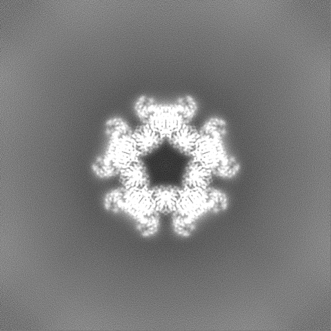
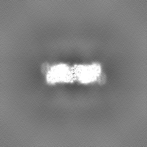
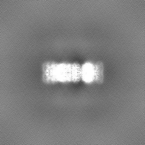
 Map data Map data | SelA sharpened map (B-factor = 92.5 A^2) | ||||||||||||||||||
 Sample Sample |
| ||||||||||||||||||
 Keywords Keywords | Selenocysteine / tRNASec / Sec-synthase / RNA BINDING PROTEIN | ||||||||||||||||||
| Function / homology |  Function and homology information Function and homology informationL-seryl-tRNASec selenium transferase / L-seryl-tRNA(Sec) selenium transferase activity / conversion of seryl-tRNAsec to selenocys-tRNAsec / L-selenocysteine biosynthetic process / selenocysteine incorporation / pyridoxal phosphate binding / identical protein binding / cytosol Similarity search - Function | ||||||||||||||||||
| Biological species |  | ||||||||||||||||||
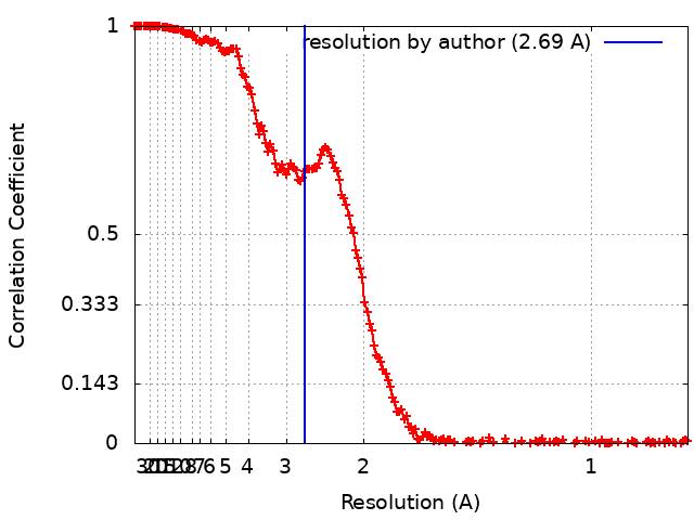
| Method | single particle reconstruction / cryo EM / Resolution: 2.69 Å | ||||||||||||||||||
 Authors Authors | Balasco Serrao VH / Minari K / Pereira HM / Thiemann OH | ||||||||||||||||||
| Funding support |  Brazil, Brazil,  United States, 5 items United States, 5 items
| ||||||||||||||||||
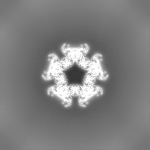
 Citation Citation | Journal: Curr Res Struct Biol / Year: 2024 Title: Bacterial selenocysteine synthase structure revealed by single-particle cryoEM. Authors: Vitor Hugo Balasco Serrão / Karine Minari / Humberto D'Muniz Pereira / Otavio Henrique Thiemann / Abstract: The 21st amino acid, selenocysteine (Sec), is synthesized on its dedicated transfer RNA (tRNA). In bacteria, Sec is synthesized from Ser-tRNA by Selenocysteine Synthase (SelA), which is a pivotal ...The 21st amino acid, selenocysteine (Sec), is synthesized on its dedicated transfer RNA (tRNA). In bacteria, Sec is synthesized from Ser-tRNA by Selenocysteine Synthase (SelA), which is a pivotal enzyme in the biosynthesis of Sec. The structural characterization of bacterial SelA is of paramount importance to decipher its catalytic mechanism and its role in the regulation of the Sec-synthesis pathway. Here, we present a comprehensive single-particle cryo-electron microscopy (SPA cryoEM) structure of the bacterial SelA with an overall resolution of 2.69 Å. Using recombinant SelA, we purified and prepared samples for single-particle cryoEM. The structural insights from SelA, combined with previous and knowledge, underscore the indispensable role of decamerization in SelA's function. Moreover, our structural analysis corroborates previous results that show that SelA adopts a pentamer of dimers configuration, and the active site architecture, substrate binding pocket, and key K295 catalytic residue are identified and described in detail. The differences in protein architecture and substrate coordination between the bacterial enzyme and its counterparts offer compelling structural evidence supporting the independent molecular evolution of the bacterial and archaea/eukarya Ser-Sec biosynthesis present in the natural world. | ||||||||||||||||||
| History |
|
- Structure visualization
| Supplemental images |
|---|

- Downloads & links
-EMDB archive
| Map data | emd_42845.map.gz | 398.5 MB | EMDB map data format | |
|---|---|---|---|---|
| Header (meta data) | emd-42845-v30.xmlemd-42845.xml | 19.2 KB 19.2 KB | Display Display | EMDB header |
| FSC (resolution estimation) | emd_42845_fsc.xml | 21.8 KB | Display | FSC data file |
| Images | 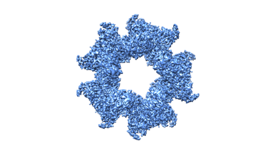 emd_42845.png emd_42845.png | 54.5 KB | ||
| Filedesc metadata | emd-42845.cif.gz | 6.6 KB | ||
| Others | emd_42845_half_map_1.map.gzemd_42845_half_map_2.map.gz | 391.8 MB 391.8 MB | ||
| Archive directory |  http://ftp.pdbj.org/pub/emdb/structures/EMD-42845ftp://ftp.pdbj.org/pub/emdb/structures/EMD-42845 http://ftp.pdbj.org/pub/emdb/structures/EMD-42845ftp://ftp.pdbj.org/pub/emdb/structures/EMD-42845 | HTTPS FTP |
-Related structure data
| Related structure data | 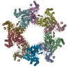 8uzwMC M: atomic model generated by this map C: citing same article ( |
|---|---|
| Similar structure data |
-Links
| EMDB pages | EMDB (EBI/PDBe) / EMDataResource |
|---|
-Map
| File | Download / File: emd_42845.map.gz / Format: CCP4 / Size: 421.9 MB / Type: IMAGE STORED AS FLOATING POINT NUMBER (4 BYTES) | ||||||||||||||||||||||||||||||||||||
|---|---|---|---|---|---|---|---|---|---|---|---|---|---|---|---|---|---|---|---|---|---|---|---|---|---|---|---|---|---|---|---|---|---|---|---|---|---|
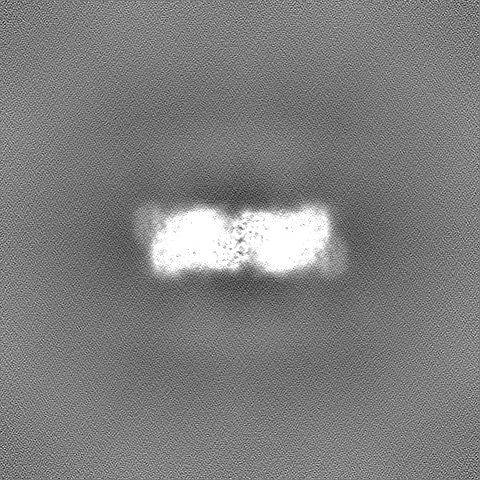
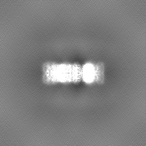
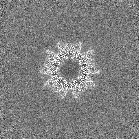
| Annotation | SelA sharpened map (B-factor = 92.5 A^2) | ||||||||||||||||||||||||||||||||||||
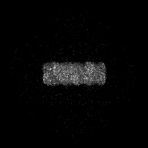
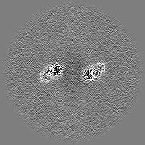
| Projections & slices | Image control
Images are generated by Spider. | ||||||||||||||||||||||||||||||||||||
| Voxel size | X=Y=Z: 0.826 Å | ||||||||||||||||||||||||||||||||||||
| Density |
| ||||||||||||||||||||||||||||||||||||
| Symmetry | Space group: 1 | ||||||||||||||||||||||||||||||||||||
| Details | EMDB XML:
|
 Z (Sec.)
 Y (Row.)
 X (Col.)


-Supplemental data
-Half map: Half map B
| File | emd_42845_half_map_1.map | ||||||||||||
|---|---|---|---|---|---|---|---|---|---|---|---|---|---|
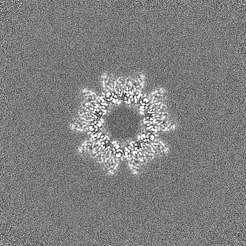
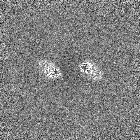
| Annotation | Half map B | ||||||||||||
| Projections & Slices |
| ||||||||||||
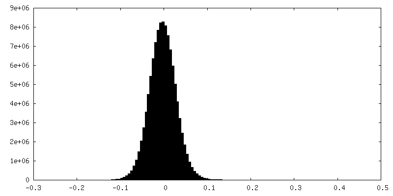
| Density Histograms |


-Half map: Half map A
| File | emd_42845_half_map_2.map | ||||||||||||
|---|---|---|---|---|---|---|---|---|---|---|---|---|---|
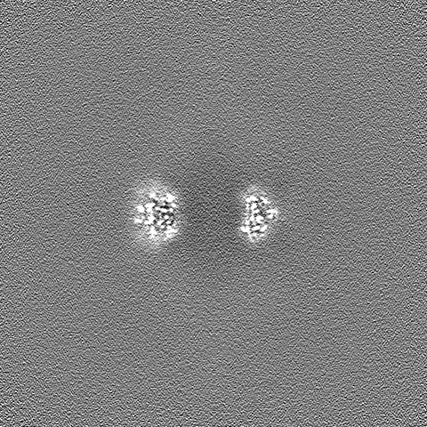
| Annotation | Half map A | ||||||||||||
| Projections & Slices |
| ||||||||||||
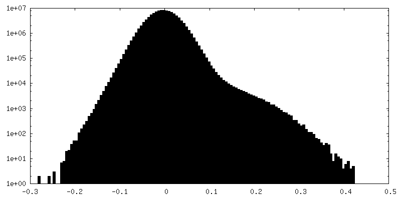
| Density Histograms |
- Sample components
-Entire : Bacterial Selenocysteine Synthase
| Entire | Name: Bacterial Selenocysteine Synthase |
|---|---|
| Components |
|
-Supramolecule #1: Bacterial Selenocysteine Synthase
| Supramolecule | Name: Bacterial Selenocysteine Synthase / type: complex / ID: 1 / Parent: 0 / Macromolecule list: all Details: Heterologously expressed E. coli homodecameric Selenocysteine Synthase |
|---|---|
| Source (natural) | Organism: |
| Molecular weight | Theoretical: 510 KDa |
-Macromolecule #1: L-seryl-tRNA(Sec) selenium transferase
| Macromolecule | Name: L-seryl-tRNA(Sec) selenium transferase / type: protein_or_peptide / ID: 1 / Number of copies: 10 / Enantiomer: LEVO / EC number: L-seryl-tRNASec selenium transferase |
|---|---|
| Source (natural) | Organism: |
| Molecular weight | Theoretical: 50.895172 KDa |
| Recombinant expression | Organism: |
| Sequence | String: MTTETRSLYS QLPAIDRLLR DSSFLSLRDT YGHTRVVELL RQMLDEAREV IRGSQTLPAW CENWAQEVDA RLTKEAQSAL RPVINLTGT VLHTNLGRAL QAEAAVEAVA QAMRSPVTLE YDLDDAGRGH RDRALAQLLC RITGAEDACI VNNNAAAVLL M LAATASGK ...String: MTTETRSLYS QLPAIDRLLR DSSFLSLRDT YGHTRVVELL RQMLDEAREV IRGSQTLPAW CENWAQEVDA RLTKEAQSAL RPVINLTGT VLHTNLGRAL QAEAAVEAVA QAMRSPVTLE YDLDDAGRGH RDRALAQLLC RITGAEDACI VNNNAAAVLL M LAATASGK EVVVSRGELV EIGGAFRIPD VMRQAGCTLH EVGTTNRTHA NDYRQAVNEN TALLMKVHTS NYSIQGFTKA ID EAELVAL GKELDVPVVT DLGSGSLVDL SQYGLPKEPM PQELIAAGVS LVSFSGD(LLP)LL GGPQAGIIVG KKEMIARLQ SHPLKRALRA DKMTLAALEA TLRLYLHPEA LSEKLPTLRL LTRSAEVIQI QAQRLQAPLA AHYGAEFAVQ VMPCLSQIGS GSLPVDRLP SAALTFTPHD GRGSHLESLA ARWRELPVPV IGRIYDGRLW LDLRCLEDEQ RFLEMLLK UniProtKB: L-seryl-tRNA(Sec) selenium transferase |
-Experimental details
-Structure determination
| Method | cryo EM |
|---|---|
 Processing Processing | single particle reconstruction |
| Aggregation state | particle |
-Sample preparation
| Concentration | 3.9 mg/mL | |||||||||||||||
|---|---|---|---|---|---|---|---|---|---|---|---|---|---|---|---|---|
| Buffer | pH: 7.5 Component:
Details: 20 mM potassium phosphate pH 7.5, 100 mM sodium chloride, 2 mM 2-mercaptoethanol, and 10 uM pyridoxal 5-phosphate. | |||||||||||||||
| Grid | Model: Quantifoil / Material: COPPER / Mesh: 400 / Support film - Material: CARBON / Support film - topology: HOLEY ARRAY / Support film - Film thickness: 20 / Pretreatment - Type: GLOW DISCHARGE / Pretreatment - Time: 20 sec. / Pretreatment - Pressure: 0.00035999999999999997 kPa | |||||||||||||||
| Vitrification | Cryogen name: ETHANE / Chamber humidity: 100 % / Chamber temperature: 295 K / Instrument: FEI VITROBOT MARK IV Details: The sample was blotted for 2.5 seconds using a force of -10 and then swiftly plunged frozen into liquid ethane using the Vitrobot Mark IV - Thermo Fisher Scientific.. | |||||||||||||||
| Details | Monodisperse sample after size-exclusion chromatography |
- Electron microscopy
| Microscope | FEI TITAN KRIOS |
|---|---|
| Image recording | Film or detector model: FEI FALCON IV (4k x 4k) / Number grids imaged: 1 / Number real images: 4592 / Average electron dose: 45.4 e/Å2 |
| Electron beam | Acceleration voltage: 300 kV / Electron source:  FIELD EMISSION GUN FIELD EMISSION GUN |
| Electron optics | Illumination mode: FLOOD BEAM / Imaging mode: BRIGHT FIELD / Nominal defocus max: 2.8000000000000003 µm / Nominal defocus min: 0.6 µm |
| Experimental equipment |  Model: Titan Krios / Image courtesy: FEI Company |
+Image processing
-Atomic model buiding 1
| Refinement | Space: REAL / Protocol: RIGID BODY FIT / Overall B value: 95.2 |
|---|---|
| Output model | PDB-8uzw: |
