 Movie
Movie Controller

[English] 日本語
 Yorodumi
- PDB-8uy1: Methylenetetrahydrofolate reductase from Chaetomium thermophilum ... -
+ Open data

- Basic information
| Entry | Database: PDB / ID: 8uy1 | ||||||
|---|---|---|---|---|---|---|---|
| Title | Methylenetetrahydrofolate reductase from Chaetomium thermophilum DSM 1495, Active (R) State | ||||||
 Components Components | Methylenetetrahydrofolate reductase-like protein | ||||||
 Keywords Keywords | OXIDOREDUCTASE / methylenetetrahydrofolate reductase / NADPH activity / oxidoreductase activity / acting on the CH-NH group of donors / NAD or NADP as acceptor cobalamin binding / one-carbon metabolism | ||||||
| Function / homology |  Function and homology information Function and homology informationmethylenetetrahydrofolate reductase [NAD(P)H] activity / : / tetrahydrofolate interconversion / FAD binding / cytosol Similarity search - Function | ||||||
| Biological species |  Thermochaetoides thermophila DSM 1495 (fungus) Thermochaetoides thermophila DSM 1495 (fungus) | ||||||
| Method |  X-RAY DIFFRACTION / SYNCHROTRON / MOLECULAR REPLACEMENT / Resolution: 3.49 Å X-RAY DIFFRACTION / SYNCHROTRON / MOLECULAR REPLACEMENT / Resolution: 3.49 Å | ||||||
 Authors Authors | Yamada, K. / Mendoza, J. / Koutmos, M. | ||||||
| Funding support |  United States, 1items United States, 1items
| ||||||
 Citation Citation | Journal: Nat Commun / Year: 2024 Title: Structural basis of S-adenosylmethionine-dependent allosteric transition from active to inactive states in methylenetetrahydrofolate reductase. Authors: Yamada, K. / Mendoza, J. / Koutmos, M. | ||||||
| History |
|
- Structure visualization
| Structure viewer | Molecule: MolmilJmol/JSmol |
|---|
- Downloads & links
-Download
| PDBx/mmCIF format | 8uy1.cif.gz | 983.7 KB | Display | PDBx/mmCIF format |
|---|---|---|---|---|
| PDB format | pdb8uy1.ent.gz | 828.2 KB | Display | PDB format |
| PDBx/mmJSON format | 8uy1.json.gz | Tree view | PDBx/mmJSON format | |
| Others |  Other downloads Other downloads |
-Validation report
| Arichive directory | https://data.pdbj.org/pub/pdb/validation_reports/uy/8uy1ftp://data.pdbj.org/pub/pdb/validation_reports/uy/8uy1 | HTTPS FTP |
|---|
-Related structure data
-Links
 PDBj
PDBj- Assembly
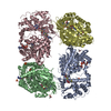
| Deposited unit | 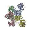
| ||||||||
|---|---|---|---|---|---|---|---|---|---|
| 1 |
| ||||||||
| Unit cell |
|
-Components
| #1: Protein | Mass: 70407.742 Da / Num. of mol.: 4 / Mutation: E21Q,L393M,V516F Source method: isolated from a genetically manipulated source Source: (gene. exp.) Thermochaetoides thermophila DSM 1495 (fungus)Strain: DSM 1495 / CBS 144.50 / IMI 039719 / Gene: CTHT_0033700 / Production host:  #2: Chemical | ChemComp-FAD /   Mass: 785.550 Da / Num. of mol.: 4 / Source method: obtained synthetically / Formula: C27H33N9O15P2 / Feature type: SUBJECT OF INVESTIGATION / Comment: FAD*YM Mass: 785.550 Da / Num. of mol.: 4 / Source method: obtained synthetically / Formula: C27H33N9O15P2 / Feature type: SUBJECT OF INVESTIGATION / Comment: FAD*YM#3: Water | ChemComp-HOH / |  Mass: 18.015 Da / Num. of mol.: 4 / Source method: isolated from a natural source / Formula: H2O Mass: 18.015 Da / Num. of mol.: 4 / Source method: isolated from a natural source / Formula: H2OHas ligand of interest | Y | |
|---|
-Experimental details
-Experiment
| Experiment | Method: X-RAY DIFFRACTION / Number of used crystals: 1 |
|---|
- Sample preparation
| Crystal | Density Matthews: 3 Å3/Da / Density % sol: 58.93 % |
|---|---|
| Crystal grow | Temperature: 293.15 K / Method: vapor diffusion, sitting drop / pH: 7.5 Details: 1-1 ratio protein to reservoir solution Protein: 25 mM Tris, pH 7.4, 50 mM potassium chloride, 500 uM FAD, and 1 mM TCEP Reservoir Solution: 0.1 M HEPES, pH 7.5, 0.1 mM potassium chloride, ...Details: 1-1 ratio protein to reservoir solution Protein: 25 mM Tris, pH 7.4, 50 mM potassium chloride, 500 uM FAD, and 1 mM TCEP Reservoir Solution: 0.1 M HEPES, pH 7.5, 0.1 mM potassium chloride, 20 mM magnesium chloride, 22% poly(acrylic acid sodium salt) 5,100 |
-Data collection
| Diffraction | Mean temperature: 100 K / Serial crystal experiment: N |
|---|---|
| Diffraction source | Source: SYNCHROTRON / Site: APS / Beamline: 21-ID-D / Wavelength: 1.1271 Å |
| Detector | Type: DECTRIS EIGER X 9M / Detector: PIXEL / Date: Jun 30, 2022 |
| Radiation | Protocol: SINGLE WAVELENGTH / Monochromatic (M) / Laue (L): M / Scattering type: x-ray |
| Radiation wavelength | Wavelength: 1.1271 Å / Relative weight: 1 |
| Reflection | Resolution: 3.41→52.04 Å / Num. obs: 46407 / % possible obs: 99.5 % / Redundancy: 6.9 % / CC1/2: 0.996 / Rmerge(I) obs: 0.164 / Rpim(I) all: 0.067 / Rrim(I) all: 0.177 / Χ2: 0.92 / Net I/σ(I): 7.7 |
| Reflection shell | Resolution: 3.41→3.53 Å / % possible obs: 99.7 % / Redundancy: 7.1 % / Rmerge(I) obs: 2.805 / Num. measured all: 32082 / Num. unique obs: 4499 / CC1/2: 0.466 / Rpim(I) all: 1.122 / Rrim(I) all: 3.024 / Χ2: 0.82 / Net I/σ(I) obs: 0.9 |
- Processing
| Software |
| ||||||||||||||||||||||||||||||||||||||||||||||||||||||||||||||||||||||||||||||||||||||||||||||||||||||||||||||||||||||||||||||||||||||||||||||||||||||||||||||||||||||||||||||||||||||
|---|---|---|---|---|---|---|---|---|---|---|---|---|---|---|---|---|---|---|---|---|---|---|---|---|---|---|---|---|---|---|---|---|---|---|---|---|---|---|---|---|---|---|---|---|---|---|---|---|---|---|---|---|---|---|---|---|---|---|---|---|---|---|---|---|---|---|---|---|---|---|---|---|---|---|---|---|---|---|---|---|---|---|---|---|---|---|---|---|---|---|---|---|---|---|---|---|---|---|---|---|---|---|---|---|---|---|---|---|---|---|---|---|---|---|---|---|---|---|---|---|---|---|---|---|---|---|---|---|---|---|---|---|---|---|---|---|---|---|---|---|---|---|---|---|---|---|---|---|---|---|---|---|---|---|---|---|---|---|---|---|---|---|---|---|---|---|---|---|---|---|---|---|---|---|---|---|---|---|---|---|---|---|---|
| Refinement | Method to determine structure: MOLECULAR REPLACEMENT / Resolution: 3.49→52.04 Å / Cor.coef. Fo:Fc: 0.906 / Cor.coef. Fo:Fc free: 0.914 / SU B: 82.341 / SU ML: 0.527 / Cross valid method: THROUGHOUT / ESU R Free: 0.623 / Stereochemistry target values: MAXIMUM LIKELIHOOD / Details: HYDROGENS HAVE BEEN USED IF PRESENT IN THE INPUT
| ||||||||||||||||||||||||||||||||||||||||||||||||||||||||||||||||||||||||||||||||||||||||||||||||||||||||||||||||||||||||||||||||||||||||||||||||||||||||||||||||||||||||||||||||||||||
| Solvent computation | Ion probe radii: 0.8 Å / Shrinkage radii: 0.8 Å / VDW probe radii: 1.2 Å / Solvent model: MASK | ||||||||||||||||||||||||||||||||||||||||||||||||||||||||||||||||||||||||||||||||||||||||||||||||||||||||||||||||||||||||||||||||||||||||||||||||||||||||||||||||||||||||||||||||||||||
| Displacement parameters | Biso mean: 157.458 Å2
| ||||||||||||||||||||||||||||||||||||||||||||||||||||||||||||||||||||||||||||||||||||||||||||||||||||||||||||||||||||||||||||||||||||||||||||||||||||||||||||||||||||||||||||||||||||||
| Refinement step | Cycle: 1 / Resolution: 3.49→52.04 Å
| ||||||||||||||||||||||||||||||||||||||||||||||||||||||||||||||||||||||||||||||||||||||||||||||||||||||||||||||||||||||||||||||||||||||||||||||||||||||||||||||||||||||||||||||||||||||
| Refine LS restraints |
|