 Movie
Movie Controller


+ Open data

- Basic information
| Entry | Database: PDB / ID: 8uvv | ||||||
|---|---|---|---|---|---|---|---|
| Title | The NanJ sialidase catalytic domain in complex with Neu5Ac | ||||||
 Components Components | Exo-alpha-sialidase NanJ | ||||||
 Keywords Keywords | HYDROLASE / Sialidase / sialic acid | ||||||
| Function / homology | N-acetyl-alpha-neuraminic acid Function and homology information Function and homology information | ||||||
| Biological species |   Clostridium perfringens (bacteria) Clostridium perfringens (bacteria) | ||||||
| Method |  X-RAY DIFFRACTION / MOLECULAR REPLACEMENT / Resolution: 2.5 Å X-RAY DIFFRACTION / MOLECULAR REPLACEMENT / Resolution: 2.5 Å | ||||||
 Authors Authors | Medley, B.J. / Boraston, A.B. | ||||||
| Funding support |  Canada, 1items Canada, 1items
| ||||||
 Citation Citation | Journal: J.Biol.Chem. / Year: 2024 Title: A "terminal" case of glycan catabolism: Structural and enzymatic characterization of the sialidases of Clostridium perfringens. Authors: Medley, B.J. / Low, K.E. / Irungu, J.D.W. / Kipchumba, L. / Daneshgar, P. / Liu, L. / Garber, J.M. / Klassen, L. / Inglis, G.D. / Boons, G.J. / Zandberg, W.F. / Abbott, D.W. / Boraston, A.B. | ||||||
| History |
|
- Structure visualization
| Structure viewer | Molecule: MolmilJmol/JSmol |
|---|
- Downloads & links
-Download
| PDBx/mmCIF format | 8uvv.cif.gz | 126.6 KB | Display | PDBx/mmCIF format |
|---|---|---|---|---|
| PDB format | pdb8uvv.ent.gz | 77.7 KB | Display | PDB format |
| PDBx/mmJSON format | 8uvv.json.gz | Tree view | PDBx/mmJSON format | |
| Others |  Other downloads Other downloads |
-Validation report
| Arichive directory | https://data.pdbj.org/pub/pdb/validation_reports/uv/8uvvftp://data.pdbj.org/pub/pdb/validation_reports/uv/8uvv | HTTPS FTP |
|---|
-Related structure data
| Related structure data | 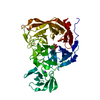 8u2aC 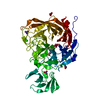 8u5oC 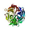 8ub5C 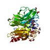 8ul7C 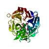 8uleC 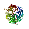 8um0C  8urlC  9c20C C: citing same article ( |
|---|---|
| Similar structure data |
-Links
 PDBj
PDBj
- Assembly
| Deposited unit | 
| ||||||||||
|---|---|---|---|---|---|---|---|---|---|---|---|
| 1 |
| ||||||||||
| Unit cell |
| ||||||||||
| Components on special symmetry positions |
|
-Components
| #1: Protein | Mass: 50047.965 Da / Num. of mol.: 1 Source method: isolated from a genetically manipulated source Source: (gene. exp.) Clostridium perfringens (bacteria) / Gene: nanJ / Production host: | ||||||||
|---|---|---|---|---|---|---|---|---|---|
| #2: Chemical |   Mass: 35.453 Da / Num. of mol.: 2 / Source method: obtained synthetically / Formula: Cl Mass: 35.453 Da / Num. of mol.: 2 / Source method: obtained synthetically / Formula: Cl#3: Sugar | ChemComp-SIA / |   Type: D-saccharide, alpha linking / Mass: 309.270 Da / Num. of mol.: 1 / Source method: obtained synthetically / Formula: C11H19NO9 / Feature type: SUBJECT OF INVESTIGATION Type: D-saccharide, alpha linking / Mass: 309.270 Da / Num. of mol.: 1 / Source method: obtained synthetically / Formula: C11H19NO9 / Feature type: SUBJECT OF INVESTIGATION#4: Water | ChemComp-HOH / |  Mass: 18.015 Da / Num. of mol.: 138 / Source method: isolated from a natural source / Formula: H2O Mass: 18.015 Da / Num. of mol.: 138 / Source method: isolated from a natural source / Formula: H2OHas ligand of interest | Y | Has protein modification | N | |
-Experimental details
-Experiment
| Experiment | Method: X-RAY DIFFRACTION / Number of used crystals: 1 |
|---|
- Sample preparation
| Crystal | Density Matthews: 3.92 Å3/Da / Density % sol: 68.63 % |
|---|---|
| Crystal grow | Temperature: 291 K / Method: vapor diffusion, sitting drop / Details: 0.1M HEPES pH 7.5, 2.0M (NH4)2S04 |
-Data collection
| Diffraction | Mean temperature: 100 K / Serial crystal experiment: N |
|---|---|
| Diffraction source | Source: ROTATING ANODE / Type: RIGAKU MICROMAX-002 / Wavelength: 1.54 Å |
| Detector | Type: DECTRIS PILATUS 200K / Detector: PIXEL / Date: Jan 20, 2022 |
| Radiation | Protocol: SINGLE WAVELENGTH / Monochromatic (M) / Laue (L): M / Scattering type: x-ray |
| Radiation wavelength | Wavelength: 1.54 Å / Relative weight: 1 |
| Reflection | Resolution: 2.5→20 Å / Num. obs: 28058 / % possible obs: 99.7 % / Redundancy: 5.5 % / Biso Wilson estimate: 42.09 Å2 / CC1/2: 0.997 / Net I/σ(I): 19 |
| Reflection shell | Resolution: 2.5→2.6 Å / Num. unique obs: 28058 / CC1/2: 0.878 |
- Processing
| Software |
| ||||||||||||||||||||||||||||||||||||||||||||||||||||||||||||||||||||||
|---|---|---|---|---|---|---|---|---|---|---|---|---|---|---|---|---|---|---|---|---|---|---|---|---|---|---|---|---|---|---|---|---|---|---|---|---|---|---|---|---|---|---|---|---|---|---|---|---|---|---|---|---|---|---|---|---|---|---|---|---|---|---|---|---|---|---|---|---|---|---|---|
| Refinement | Method to determine structure: MOLECULAR REPLACEMENT / Resolution: 2.5→19.67 Å / SU ML: 0.344 / Cross valid method: FREE R-VALUE / σ(F): 1.33 / Phase error: 27.4477 Stereochemistry target values: GeoStd + Monomer Library + CDL v1.2
| ||||||||||||||||||||||||||||||||||||||||||||||||||||||||||||||||||||||
| Solvent computation | Shrinkage radii: 0.9 Å / VDW probe radii: 1.1 Å / Solvent model: FLAT BULK SOLVENT MODEL | ||||||||||||||||||||||||||||||||||||||||||||||||||||||||||||||||||||||
| Displacement parameters | Biso mean: 42.76 Å2 | ||||||||||||||||||||||||||||||||||||||||||||||||||||||||||||||||||||||
| Refinement step | Cycle: LAST / Resolution: 2.5→19.67 Å
| ||||||||||||||||||||||||||||||||||||||||||||||||||||||||||||||||||||||
| Refine LS restraints |
| ||||||||||||||||||||||||||||||||||||||||||||||||||||||||||||||||||||||
| LS refinement shell |
|
