 Movie
Movie Controller


+ Open data

- Basic information
| Entry | Database: PDB / ID: 8um0 | ||||||
|---|---|---|---|---|---|---|---|
| Title | The structure of NanH in complex with Neu5,7,9Ac(2,6)-LAcNAc | ||||||
 Components Components | Sialidase | ||||||
 Keywords Keywords | HYDROLASE / Sialidase | ||||||
| Function / homology |  Function and homology information Function and homology informationganglioside catabolic process / oligosaccharide catabolic process / exo-alpha-sialidase / exo-alpha-sialidase activity / membrane / cytoplasm Similarity search - Function | ||||||
| Biological species |  Clostridium perfringens ATCC 13124 (bacteria) Clostridium perfringens ATCC 13124 (bacteria) | ||||||
| Method |  X-RAY DIFFRACTION / MOLECULAR REPLACEMENT / Resolution: 2.46 Å X-RAY DIFFRACTION / MOLECULAR REPLACEMENT / Resolution: 2.46 Å | ||||||
 Authors Authors | Medley, B.J. / Boraston, A.B. | ||||||
| Funding support |  Canada, 1items Canada, 1items
| ||||||
 Citation Citation | Journal: J.Biol.Chem. / Year: 2024 Title: A "terminal" case of glycan catabolism: Structural and enzymatic characterization of the sialidases of Clostridium perfringens. Authors: Medley, B.J. / Low, K.E. / Irungu, J.D.W. / Kipchumba, L. / Daneshgar, P. / Liu, L. / Garber, J.M. / Klassen, L. / Inglis, G.D. / Boons, G.J. / Zandberg, W.F. / Abbott, D.W. / Boraston, A.B. | ||||||
| History |
|
- Structure visualization
| Structure viewer | Molecule: MolmilJmol/JSmol |
|---|
- Downloads & links
-Download
| PDBx/mmCIF format | 8um0.cif.gz | 90.7 KB | Display | PDBx/mmCIF format |
|---|---|---|---|---|
| PDB format | pdb8um0.ent.gz | 65.4 KB | Display | PDB format |
| PDBx/mmJSON format | 8um0.json.gz | Tree view | PDBx/mmJSON format | |
| Others |  Other downloads Other downloads |
-Validation report
| Arichive directory | https://data.pdbj.org/pub/pdb/validation_reports/um/8um0ftp://data.pdbj.org/pub/pdb/validation_reports/um/8um0 | HTTPS FTP |
|---|
-Related structure data
| Related structure data | 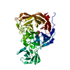 8u2aC 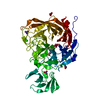 8u5oC 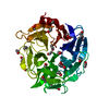 8ub5C 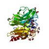 8ul7C 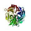 8uleC  8urlC  8uvvC  9c20C C: citing same article ( |
|---|---|
| Similar structure data |
-Links
 PDBj
PDBj

- Assembly
| Deposited unit | 
| ||||||||
|---|---|---|---|---|---|---|---|---|---|
| 1 |
| ||||||||
| Unit cell |
|
-Components
| #1: Protein | Mass: 43065.930 Da / Num. of mol.: 1 Source method: isolated from a genetically manipulated source Source: (gene. exp.) Clostridium perfringens ATCC 13124 (bacteria)Gene: nanH / Production host: | ||||||
|---|---|---|---|---|---|---|---|
| #2: Polysaccharide | beta-D-galactopyranose-(1-4)-2-acetamido-2-deoxy-beta-D-glucopyranose Source method: isolated from a genetically manipulated source | ||||||
| #3: Chemical | ChemComp-X1F / Mass: 393.343 Da / Num. of mol.: 1 / Source method: obtained synthetically / Formula: C15H23NO11 | ||||||
| #4: Chemical | ChemComp-EDO /   Mass: 62.068 Da / Num. of mol.: 10 / Source method: obtained synthetically / Formula: C2H6O2 Mass: 62.068 Da / Num. of mol.: 10 / Source method: obtained synthetically / Formula: C2H6O2#5: Water | ChemComp-HOH / |  Mass: 18.015 Da / Num. of mol.: 129 / Source method: isolated from a natural source / Formula: H2O Mass: 18.015 Da / Num. of mol.: 129 / Source method: isolated from a natural source / Formula: H2OHas ligand of interest | Y | Has protein modification | N | |
-Experimental details
-Experiment
| Experiment | Method: X-RAY DIFFRACTION / Number of used crystals: 1 |
|---|
- Sample preparation
| Crystal | Density Matthews: 2.24 Å3/Da / Density % sol: 45.05 % |
|---|---|
| Crystal grow | Temperature: 291 K / Method: vapor diffusion, hanging drop / Details: 0.1 M HEPES:NaOH pH 7.5, 20% PEG 8000 |
-Data collection
| Diffraction | Mean temperature: 100 K / Serial crystal experiment: N |
|---|---|
| Diffraction source | Source: ROTATING ANODE / Type: RIGAKU MICROMAX-002 / Wavelength: 1.54 Å |
| Detector | Type: DECTRIS PILATUS 200K / Detector: PIXEL / Date: Jan 30, 2021 |
| Radiation | Protocol: SINGLE WAVELENGTH / Monochromatic (M) / Laue (L): M / Scattering type: x-ray |
| Radiation wavelength | Wavelength: 1.54 Å / Relative weight: 1 |
| Reflection | Resolution: 2.46→24 Å / Num. obs: 25987 / % possible obs: 99.3 % / Redundancy: 3.9 % / CC1/2: 0.993 / Net I/σ(I): 16.1 |
| Reflection shell | Resolution: 2.46→2.65 Å / Num. unique obs: 1267 / CC1/2: 0.815 |
- Processing
| Software |
| ||||||||||||||||||||||||||||||||||||||||||
|---|---|---|---|---|---|---|---|---|---|---|---|---|---|---|---|---|---|---|---|---|---|---|---|---|---|---|---|---|---|---|---|---|---|---|---|---|---|---|---|---|---|---|---|
| Refinement | Method to determine structure: MOLECULAR REPLACEMENT / Resolution: 2.46→23.8 Å / SU ML: 0.31 / Cross valid method: FREE R-VALUE / σ(F): 1.35 / Phase error: 24.47 / Stereochemistry target values: ML
| ||||||||||||||||||||||||||||||||||||||||||
| Solvent computation | Shrinkage radii: 0.9 Å / VDW probe radii: 1.1 Å / Solvent model: FLAT BULK SOLVENT MODEL | ||||||||||||||||||||||||||||||||||||||||||
| Refinement step | Cycle: LAST / Resolution: 2.46→23.8 Å
| ||||||||||||||||||||||||||||||||||||||||||
| Refine LS restraints |
| ||||||||||||||||||||||||||||||||||||||||||
| LS refinement shell |
|
