Movie
Movie Controller


+ Open data

- Basic information
| Entry | Database: PDB / ID: 8uvh | |||||||||||||||
|---|---|---|---|---|---|---|---|---|---|---|---|---|---|---|---|---|
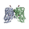
| Title | Structure of NaCT-succ complex | |||||||||||||||
 Components Components | Solute carrier family 13 member 5 | |||||||||||||||
 Keywords Keywords | TRANSPORT PROTEIN / Na(+)/citrate cotransporter(NaCT) / Solute carries / Elevator type alternating access / membrane protein | |||||||||||||||
| Function / homology |  Function and homology information Function and homology informationorganic acid:sodium symporter activity / fumarate transport / oxaloacetate transport / succinate transport / sodium:dicarboxylate symporter activity / citrate transmembrane transporter activity / citrate transport / Sodium-coupled sulphate, di- and tri-carboxylate transporters / alpha-ketoglutarate transport / succinate transmembrane transporter activity ...organic acid:sodium symporter activity / fumarate transport / oxaloacetate transport / succinate transport / sodium:dicarboxylate symporter activity / citrate transmembrane transporter activity / citrate transport / Sodium-coupled sulphate, di- and tri-carboxylate transporters / alpha-ketoglutarate transport / succinate transmembrane transporter activity / cellular response to lithium ion / transmembrane transport / nucleoplasm / identical protein binding / plasma membrane / cytosol Similarity search - Function | |||||||||||||||
| Biological species |  Homo sapiens (human) Homo sapiens (human) | |||||||||||||||
| Method | ELECTRON MICROSCOPY / single particle reconstruction / cryo EM / Resolution: 2.33 Å | |||||||||||||||
 Authors Authors | Li, Y. / Wang, D.N. / Mindell, J.A. / Rice, W.J. / Song, J. / Mikusevic, V. / Marden, J.J. / Becerril, A. / Kuang, H. / Wang, B. | |||||||||||||||
| Funding support |  United States, 4items United States, 4items
| |||||||||||||||
 Citation Citation | Journal: Nat Struct Mol Biol / Year: 2025 Title: Substrate translocation and inhibition in human dicarboxylate transporter NaDC3. Authors: Yan Li / Jinmei Song / Vedrana Mikusevic / Jennifer J Marden / Alissa Becerril / Huihui Kuang / Bing Wang / William J Rice / Joseph A Mindell / Da-Neng Wang / Abstract: The human high-affinity sodium-dicarboxylate cotransporter (NaDC3) imports various substrates into the cell as tricarboxylate acid cycle intermediates, lipid biosynthesis precursors and signaling ...The human high-affinity sodium-dicarboxylate cotransporter (NaDC3) imports various substrates into the cell as tricarboxylate acid cycle intermediates, lipid biosynthesis precursors and signaling molecules. Understanding the cellular signaling process and developing inhibitors require knowledge of the structural basis of the dicarboxylate specificity and inhibition mechanism of NaDC3. To this end, we determined the cryo-electron microscopy structures of NaDC3 in various dimers, revealing the protomer in three conformations: outward-open C, outward-occluded C and inward-open C. A dicarboxylate is first bound and recognized in C and how the substrate interacts with NaDC3 in C likely helps to further determine the substrate specificity. A phenylalanine from the scaffold domain interacts with the bound dicarboxylate in the C state and modulates the kinetic barrier to the transport domain movement. Structural comparison of an inhibitor-bound structure of NaDC3 to that of the sodium-dependent citrate transporter suggests ways for making an inhibitor that is specific for NaDC3. | |||||||||||||||
| History |
|
- Structure visualization
| Structure viewer | Molecule: MolmilJmol/JSmol |
|---|
- Downloads & links
-Download
| PDBx/mmCIF format | 8uvh.cif.gz | 166.3 KB | Display | PDBx/mmCIF format |
|---|---|---|---|---|
| PDB format | pdb8uvh.ent.gz | 131.1 KB | Display | PDB format |
| PDBx/mmJSON format | 8uvh.json.gz | Tree view | PDBx/mmJSON format | |
| Others |  Other downloads Other downloads |
-Validation report
| Arichive directory | https://data.pdbj.org/pub/pdb/validation_reports/uv/8uvhftp://data.pdbj.org/pub/pdb/validation_reports/uv/8uvh | HTTPS FTP |
|---|
-Related structure data
| Related structure data |  42620MC  8uvbC  8uvcC 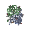 8uvdC  8uveC  8uvfC  8uvgC  8uviC M: map data used to model this data C: citing same article ( |
|---|---|
| Similar structure data |


-Links
 PDBj
PDBj- Assembly
| Deposited unit | 
|
|---|---|
| 1 |
|
-Components
| #1: Protein | Mass: 63110.812 Da / Num. of mol.: 2 Source method: isolated from a genetically manipulated source Details: Solute carrier family 13 member 5 / Source: (gene. exp.) Homo sapiens (human) / Gene: SLC13A5, NACT / Production host:  Trichoplusia ni (cabbage looper) / References: UniProt: Q86YT5 Trichoplusia ni (cabbage looper) / References: UniProt: Q86YT5Has ligand of interest | Y | Has protein modification | N | |
|---|
-Experimental details
-Experiment
| Experiment | Method: ELECTRON MICROSCOPY |
|---|---|
| EM experiment | Aggregation state: PARTICLE / 3D reconstruction method: single particle reconstruction |
- Sample preparation
| Component | Name: Dimer of NaCT complex in succinate / Type: COMPLEX / Entity ID: all / Source: RECOMBINANT | ||||||||||||||||||||
|---|---|---|---|---|---|---|---|---|---|---|---|---|---|---|---|---|---|---|---|---|---|
| Molecular weight |
| ||||||||||||||||||||
| Source (natural) | Organism: Homo sapiens (human) | ||||||||||||||||||||
| Source (recombinant) | Organism: Trichoplusia ni (cabbage looper) | ||||||||||||||||||||
| Buffer solution | pH: 7.5 | ||||||||||||||||||||
| Buffer component |
| ||||||||||||||||||||
| Specimen | Conc.: 1 mg/ml / Embedding applied: NO / Shadowing applied: NO / Staining applied: NO / Vitrification applied: YES Details: This sample was performed an amphipol(PMAL-C8) exchange at a 1:5 protein:amphipol weight ratio | ||||||||||||||||||||
| Specimen support | Details: Hold 10s before glow discharge / Grid material: GOLD / Grid mesh size: 300 divisions/in. / Grid type: UltrAuFoil R1.2/1.3 | ||||||||||||||||||||
| Vitrification | Instrument: FEI VITROBOT MARK IV / Cryogen name: ETHANE / Humidity: 100 % / Chamber temperature: 281.15 K |
- Electron microscopy imaging
| Experimental equipment |  Model: Titan Krios / Image courtesy: FEI Company |
|---|---|
| Microscopy | Model: FEI TITAN KRIOS |
| Electron gun | Electron source:  FIELD EMISSION GUN / Accelerating voltage: 300 kV / Illumination mode: FLOOD BEAM FIELD EMISSION GUN / Accelerating voltage: 300 kV / Illumination mode: FLOOD BEAM |
| Electron lens | Mode: BRIGHT FIELD / Nominal magnification: 105000 X / Calibrated magnification: 105000 X / Nominal defocus max: 1600 nm / Nominal defocus min: 1200 nm / Calibrated defocus min: 800 nm / Calibrated defocus max: 3000 nm / Cs: 2.7 mm / C2 aperture diameter: 70 µm / Alignment procedure: COMA FREE |
| Specimen holder | Cryogen: NITROGEN / Specimen holder model: FEI TITAN KRIOS AUTOGRID HOLDER / Temperature (max): 80 K / Temperature (min): 80 K / Residual tilt: 0.05 mradians |
| Image recording | Average exposure time: 2.5 sec. / Electron dose: 52.75 e/Å2 / Film or detector model: GATAN K3 BIOQUANTUM (6k x 4k) / Num. of grids imaged: 1 / Num. of real images: 10359 Details: 5868 untilted images and 4491 40 degrees tilted images were collected in super resolution mode at 50 frames per micrograph |
| EM imaging optics | Energyfilter name: GIF Bioquantum / Energyfilter slit width: 20 eV |
| Image scans | Sampling size: 5 µm / Width: 5760 / Height: 4092 |
- Processing
| EM software |
| ||||||||||||||||||||||||||||||||||||||||||||||||
|---|---|---|---|---|---|---|---|---|---|---|---|---|---|---|---|---|---|---|---|---|---|---|---|---|---|---|---|---|---|---|---|---|---|---|---|---|---|---|---|---|---|---|---|---|---|---|---|---|---|
| Image processing | Details: The micrographs with an overall resolution worse than 5 angstrom were excluded | ||||||||||||||||||||||||||||||||||||||||||||||||
| CTF correction | Details: CTF amplitude correction was performed after particle polishing Type: PHASE FLIPPING AND AMPLITUDE CORRECTION | ||||||||||||||||||||||||||||||||||||||||||||||||
| Particle selection | Num. of particles selected: 3125207 Details: Particles were selected from 5868 untilted images and 4491 40 degree tilted images | ||||||||||||||||||||||||||||||||||||||||||||||||
| Symmetry | Point symmetry: C2 (2 fold cyclic) | ||||||||||||||||||||||||||||||||||||||||||||||||
| 3D reconstruction | Resolution: 2.33 Å / Resolution method: FSC 0.143 CUT-OFF / Num. of particles: 323571 / Algorithm: FOURIER SPACE / Num. of class averages: 1 / Symmetry type: POINT | ||||||||||||||||||||||||||||||||||||||||||||||||
| Atomic model building | B value: 52.62 / Protocol: OTHER / Space: REAL / Target criteria: cross-correlation coefficient Details: Initial local fitting was done using Chimera and then coot was used for ajustment. | ||||||||||||||||||||||||||||||||||||||||||||||||
| Atomic model building | PDB-ID: 7jsk Pdb chain-ID: AB / Accession code: 7jsk / Chain residue range: 1-568 / Details: The whole model was used as an initial model / Pdb chain residue range: 1-568 / Source name: PDB / Type: experimental model |