 Movie
Movie Controller


+ Open data

- Basic information
| Entry | Database: PDB / ID: 8uqt | ||||||
|---|---|---|---|---|---|---|---|
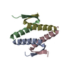
| Title | Crystal structure of the Tree Shrew p53 tetramerization domain | ||||||
 Components Components | Cellular tumor antigen p53 | ||||||
 Keywords Keywords | TRANSCRIPTION / Tree Shrew p53 tetramerization domain stability | ||||||
| Function / homology |  Function and homology information Function and homology informationprotein tetramerization / PML body / rhythmic process / transcription cis-regulatory region binding / mitochondrial matrix / cell cycle / DNA-binding transcription factor activity / apoptotic process / endoplasmic reticulum / metal ion binding Similarity search - Function | ||||||
| Biological species |  | ||||||
| Method |  X-RAY DIFFRACTION / SYNCHROTRON / MOLECULAR REPLACEMENT / Resolution: 1.16 Å X-RAY DIFFRACTION / SYNCHROTRON / MOLECULAR REPLACEMENT / Resolution: 1.16 Å | ||||||
 Authors Authors | Wahba, H.M. / Sakaguchi, S. / Nakagawa, N. / Wada, J. / Kamada, R. / Sakaguchi, K. / Omichinski, J.G. | ||||||
| Funding support |  Canada, 1items Canada, 1items
| ||||||
 Citation Citation | Journal: Int J Mol Sci / Year: 2023 Title: Highly Similar Tetramerization Domains from the p53 Protein of Different Mammalian Species Possess Varying Biophysical, Functional and Structural Properties. Authors: Sakaguchi, S. / Nakagawa, N. / Wahba, H.M. / Wada, J. / Kamada, R. / Omichinski, J.G. / Sakaguchi, K. | ||||||
| History |
|
- Structure visualization
| Structure viewer | Molecule: MolmilJmol/JSmol |
|---|
- Downloads & links
-Download
| PDBx/mmCIF format | 8uqt.cif.gz | 54.1 KB | Display | PDBx/mmCIF format |
|---|---|---|---|---|
| PDB format | pdb8uqt.ent.gz | 38.8 KB | Display | PDB format |
| PDBx/mmJSON format | 8uqt.json.gz | Tree view | PDBx/mmJSON format | |
| Others |  Other downloads Other downloads |
-Validation report
| Summary document | 8uqt_validation.pdf.gz | 739.1 KB | Display | wwPDB validaton report |
|---|---|---|---|---|
| Full document | 8uqt_full_validation.pdf.gz | 740.1 KB | Display | |
| Data in XML | 8uqt_validation.xml.gz | 6.2 KB | Display | |
| Data in CIF | 8uqt_validation.cif.gz | 7.8 KB | Display | |
| Arichive directory | https://data.pdbj.org/pub/pdb/validation_reports/uq/8uqtftp://data.pdbj.org/pub/pdb/validation_reports/uq/8uqt | HTTPS FTP |
-Related structure data

-Links
 PDBj
PDBj


- Assembly
| Deposited unit | 
| |||||||||||||||
|---|---|---|---|---|---|---|---|---|---|---|---|---|---|---|---|---|
| 1 | 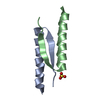
| |||||||||||||||
| Unit cell |
| |||||||||||||||
| Components on special symmetry positions |
|
-Components
| #1: Protein/peptide | Mass: 3879.402 Da / Num. of mol.: 2 Source method: isolated from a genetically manipulated source Source: (gene. exp.)  #2: Chemical | ChemComp-SO4 / |   Mass: 96.063 Da / Num. of mol.: 1 / Source method: obtained synthetically / Formula: SO4 / Feature type: SUBJECT OF INVESTIGATION Mass: 96.063 Da / Num. of mol.: 1 / Source method: obtained synthetically / Formula: SO4 / Feature type: SUBJECT OF INVESTIGATION#3: Water | ChemComp-HOH / |  Mass: 18.015 Da / Num. of mol.: 99 / Source method: isolated from a natural source / Formula: H2O Mass: 18.015 Da / Num. of mol.: 99 / Source method: isolated from a natural source / Formula: H2OHas ligand of interest | Y | |
|---|
-Experimental details
-Experiment
| Experiment | Method: X-RAY DIFFRACTION / Number of used crystals: 1 |
|---|
- Sample preparation
| Crystal | Density Matthews: 2.17 Å3/Da / Density % sol: 43.24 % |
|---|---|
| Crystal grow | Temperature: 296 K / Method: vapor diffusion, hanging drop Details: 0.6 M Lithium sulfate monohydrate, 2% PEG 8000, 10% glycerol |
-Data collection
| Diffraction | Mean temperature: 100 K / Serial crystal experiment: N |
|---|---|
| Diffraction source | Source: SYNCHROTRON / Site: CHESS  / Beamline: 7B2 / Wavelength: 1 Å / Beamline: 7B2 / Wavelength: 1 Å |
| Detector | Type: DECTRIS PILATUS 6M / Detector: PIXEL / Date: Oct 19, 2020 |
| Radiation | Protocol: SINGLE WAVELENGTH / Monochromatic (M) / Laue (L): M / Scattering type: x-ray |
| Radiation wavelength | Wavelength: 1 Å / Relative weight: 1 |
| Reflection | Resolution: 1.16→45.46 Å / Num. obs: 21880 / % possible obs: 91.7 % / Redundancy: 9.9 % / CC1/2: 1 / Net I/σ(I): 28.35 |
| Reflection shell | Resolution: 1.16→1.201 Å / Num. unique obs: 21822 / CC1/2: 0.84 |
- Processing
| Software |
| |||||||||||||||||||||||||||||||||||||||||||||||||||||||||||||||||||||||||||||||||||||||||||||||||||||||||
|---|---|---|---|---|---|---|---|---|---|---|---|---|---|---|---|---|---|---|---|---|---|---|---|---|---|---|---|---|---|---|---|---|---|---|---|---|---|---|---|---|---|---|---|---|---|---|---|---|---|---|---|---|---|---|---|---|---|---|---|---|---|---|---|---|---|---|---|---|---|---|---|---|---|---|---|---|---|---|---|---|---|---|---|---|---|---|---|---|---|---|---|---|---|---|---|---|---|---|---|---|---|---|---|---|---|---|
| Refinement | Method to determine structure: MOLECULAR REPLACEMENT / Resolution: 1.16→45.46 Å / SU ML: 0.1 / Cross valid method: FREE R-VALUE / σ(F): 1.38 / Phase error: 18.85 / Stereochemistry target values: ML
| |||||||||||||||||||||||||||||||||||||||||||||||||||||||||||||||||||||||||||||||||||||||||||||||||||||||||
| Solvent computation | Shrinkage radii: 0.9 Å / VDW probe radii: 1.11 Å / Solvent model: FLAT BULK SOLVENT MODEL | |||||||||||||||||||||||||||||||||||||||||||||||||||||||||||||||||||||||||||||||||||||||||||||||||||||||||
| Refinement step | Cycle: LAST / Resolution: 1.16→45.46 Å
| |||||||||||||||||||||||||||||||||||||||||||||||||||||||||||||||||||||||||||||||||||||||||||||||||||||||||
| Refine LS restraints |
| |||||||||||||||||||||||||||||||||||||||||||||||||||||||||||||||||||||||||||||||||||||||||||||||||||||||||
| LS refinement shell |
|
