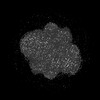
 Movie
Movie Controller

[English] 日本語
 Yorodumi
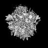
- PDB-8uop: Major interface of Streptococcal surface enolase dimer from AP53 ... -
+ Open data

- Basic information
| Entry | Database: PDB / ID: 8uop | ||||||
|---|---|---|---|---|---|---|---|
| Title | Major interface of Streptococcal surface enolase dimer from AP53 group A streptococcus bound to a lipid vesicle | ||||||
 Components Components | Enolase | ||||||
 Keywords Keywords | LYASE / Glycolysis / plasminogen binder / membrane protein | ||||||
| Function / homology |  Function and homology information Function and homology informationphosphopyruvate hydratase / phosphopyruvate hydratase complex / phosphopyruvate hydratase activity / peptidoglycan-based cell wall / glycolytic process / magnesium ion binding / cell surface / extracellular region Similarity search - Function | ||||||
| Biological species |  Streptococcus pyogenes (bacteria) Streptococcus pyogenes (bacteria) | ||||||
| Method | ELECTRON MICROSCOPY / single particle reconstruction / cryo EM / Resolution: 3.8 Å | ||||||
 Authors Authors | Tjia-Fleck, S. / Readnour, B.M. / Castellino, F.J. | ||||||
| Funding support |  United States, 1items United States, 1items
| ||||||
 Citation Citation | Journal: To Be Published Title: Streptococcus surface alpha enolase exposed dimers were found to be the active form on lipid surface that binds to human plasminogen Authors: Tjia-Fleck, S. / Readnour, B.M. / Castellino, F.J. | ||||||
| History |
|
- Structure visualization
| Structure viewer | Molecule: MolmilJmol/JSmol |
|---|
- Downloads & links
-Download
| PDBx/mmCIF format | 8uop.cif.gz | 312.3 KB | Display | PDBx/mmCIF format |
|---|---|---|---|---|
| PDB format | pdb8uop.ent.gz | 259.1 KB | Display | PDB format |
| PDBx/mmJSON format | 8uop.json.gz | Tree view | PDBx/mmJSON format | |
| Others |  Other downloads Other downloads |
-Validation report
| Arichive directory | https://data.pdbj.org/pub/pdb/validation_reports/uo/8uopftp://data.pdbj.org/pub/pdb/validation_reports/uo/8uop | HTTPS FTP |
|---|
-Related structure data
| Related structure data | 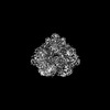 42435MC 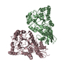 8uoyC  8uq6C M: map data used to model this data C: citing same article ( |
|---|---|
| Similar structure data |
-Links
 PDBj
PDBj
- Assembly
| Deposited unit | 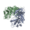
|
|---|---|
| 1 |
|
-Components
| #1: Protein | Mass: 47543.281 Da / Num. of mol.: 2 Source method: isolated from a genetically manipulated source Source: (gene. exp.) Streptococcus pyogenes (bacteria)Gene: eno, E0F66_01300, E0F67_02490, FGO82_08975, SAMEA1711581_01820, SAMEA864267_00487 Production host: References: UniProt: A3F8V6, phosphopyruvate hydratase |
|---|
-Experimental details
-Experiment
| Experiment | Method: ELECTRON MICROSCOPY |
|---|---|
| EM experiment | Aggregation state: PARTICLE / 3D reconstruction method: single particle reconstruction |
- Sample preparation
| Component | Name: Streptococcal surface enolase major interface embedded into the surface of a lipid vesicle Type: COMPLEX / Entity ID: all / Source: RECOMBINANT |
|---|---|
| Molecular weight | Value: 0.095 MDa / Experimental value: YES |
| Source (natural) | Organism: Streptococcus pyogenes (bacteria) |
| Source (recombinant) | Organism: |
| Buffer solution | pH: 7.4 |
| Specimen | Embedding applied: NO / Shadowing applied: NO / Staining applied: NO / Vitrification applied: YES |
| Specimen support | Grid material: GOLD / Grid mesh size: 400 divisions/in. |
| Vitrification | Instrument: FEI VITROBOT MARK IV / Cryogen name: ETHANE / Humidity: 100 % / Chamber temperature: 298 K |
- Electron microscopy imaging
| Experimental equipment |  Model: Titan Krios / Image courtesy: FEI Company |
|---|---|
| Microscopy | Model: FEI TITAN KRIOS |
| Electron gun | Electron source:  FIELD EMISSION GUN / Accelerating voltage: 300 kV / Illumination mode: SPOT SCAN FIELD EMISSION GUN / Accelerating voltage: 300 kV / Illumination mode: SPOT SCAN |
| Electron lens | Mode: BRIGHT FIELD / Nominal magnification: 81000 X / Nominal defocus max: 29000 nm / Nominal defocus min: 1000 nm / Cs: 2.7 mm / Alignment procedure: BASIC |
| Specimen holder | Cryogen: NITROGEN / Specimen holder model: FEI TITAN KRIOS AUTOGRID HOLDER |
| Image recording | Electron dose: 60 e/Å2 / Film or detector model: GATAN K3 (6k x 4k) |
- Processing
| EM software |
| ||||||||||||||||||
|---|---|---|---|---|---|---|---|---|---|---|---|---|---|---|---|---|---|---|---|
| CTF correction | Type: PHASE FLIPPING AND AMPLITUDE CORRECTION | ||||||||||||||||||
| Particle selection | Num. of particles selected: 3300000 | ||||||||||||||||||
| Symmetry | Point symmetry: C2 (2 fold cyclic) | ||||||||||||||||||
| 3D reconstruction | Resolution: 3.8 Å / Resolution method: FSC 0.143 CUT-OFF / Num. of particles: 525846 / Algorithm: FOURIER SPACE / Symmetry type: POINT |
