 Movie
Movie Controller


+ Open data

- Basic information
| Entry | Database: PDB / ID: 8uki | ||||||
|---|---|---|---|---|---|---|---|
| Title | Crystal structure of 04_A06 Fab | ||||||
 Components Components |
| ||||||
 Keywords Keywords | IMMUNE SYSTEM / Fab / broadly neutralizing antibody / HIV-1 / CD4 binding site | ||||||
| Biological species |  Homo sapiens (human) Homo sapiens (human) | ||||||
| Method |  X-RAY DIFFRACTION / SYNCHROTRON / MOLECULAR REPLACEMENT / Resolution: 1.75 Å X-RAY DIFFRACTION / SYNCHROTRON / MOLECULAR REPLACEMENT / Resolution: 1.75 Å | ||||||
 Authors Authors | DeLaitsch, A.T. / Gristick, H.B. / Bjorkman, P.J. | ||||||
| Funding support |  United States, 1items United States, 1items
| ||||||
 Citation Citation | Journal: Nat Immunol / Year: 2025 Title: Profiling of HIV-1 elite neutralizer cohort reveals a CD4bs bnAb for HIV-1 prevention and therapy. Authors: Lutz Gieselmann / Andrew T DeLaitsch / Malena Rohde / Henning Gruell / Christoph Kreer / Meryem Seda Ercanoglu / Harry B Gristick / Philipp Schommers / Elvin Ahmadov / Caelan Radford / ...Authors: Lutz Gieselmann / Andrew T DeLaitsch / Malena Rohde / Henning Gruell / Christoph Kreer / Meryem Seda Ercanoglu / Harry B Gristick / Philipp Schommers / Elvin Ahmadov / Caelan Radford / Andrea Mazzolini / Lily Zhang / Anthony P West / Johanna Worczinski / Anna Ashurov / Maren L Reichwein / Jacqueline Knüfer / Ricarda Stumpf / Nonhlanhla N Mkhize / Haajira Kaldine / Sinethemba Bhebhe / Sharvari Deshpande / Federico Giovannoni / Erin Stefanutti / Fabio Benigni / Colin Havenar-Daughton / Davide Corti / Arne Kroidl / Anurag Adhikari / Aubin J Nanfack / Georgia E Ambada / Ralf Duerr / Lucas Maganga / Wiston William / Nyanda E Ntinginya / Timo Wolf / Christof Geldmacher / Michael Hoelscher / Clara Lehmann / Penny L Moore / Thierry Mora / Aleksandra M Walczak / Peter B Gilbert / Nicole A Doria-Rose / Yunda Huang / Jesse D Bloom / Michael S Seaman / Pamela J Bjorkman / Florian Klein /     Abstract: Administration of HIV-1 neutralizing antibodies can suppress viremia and prevent infection in vivo. However, clinical use is challenged by envelope diversity and rapid viral escape. Here, we ...Administration of HIV-1 neutralizing antibodies can suppress viremia and prevent infection in vivo. However, clinical use is challenged by envelope diversity and rapid viral escape. Here, we performed single B cell profiling of 32 top HIV-1 elite neutralizers to identify broadly neutralizing antibodies with highest antiviral activity. From 831 expressed monoclonal antibodies, we identified 04_A06, a V1-2-encoded broadly neutralizing antibody to the CD4 binding site with remarkable breadth and potency against multiclade pseudovirus panels (geometric mean half-maximal inhibitory concentration = 0.059 µg ml, breadth = 98.5%, 332 strains). Moreover, 04_A06 was not susceptible to classic CD4 binding site escape variants and maintained full viral suppression in HIV-1-infected humanized mice. Structural analyses revealed an unusually long 11-amino-acid heavy chain insertion that facilitates interprotomer contacts with highly conserved residues on the adjacent gp120 protomer. Finally, 04_A06 demonstrated high activity against contemporaneously circulating viruses from the Antibody-Mediated Prevention trials (geometric mean half-maximal inhibitory concentration = 0.082 µg ml, breadth = 98.4%, 191 virus strains), and in silico modeling for 04_A06LS predicted prevention efficacy of >93%. Thus, 04_A06 will provide unique opportunities for effective treatment and prevention of HIV-1 infection. | ||||||
| History |
|
- Structure visualization
| Structure viewer | Molecule:  MolmilJmol/JSmol MolmilJmol/JSmol |
|---|
- Downloads & links
-Download
| PDBx/mmCIF format | 8uki.cif.gz | 184 KB | Display | PDBx/mmCIF format |
|---|---|---|---|---|
| PDB format | pdb8uki.ent.gz | 142.3 KB | Display | PDB format |
| PDBx/mmJSON format | 8uki.json.gz | Tree view | PDBx/mmJSON format | |
| Others |  Other downloads Other downloads |
-Validation report
| Arichive directory | https://data.pdbj.org/pub/pdb/validation_reports/uk/8ukiftp://data.pdbj.org/pub/pdb/validation_reports/uk/8uki | HTTPS FTP |
|---|
-Related structure data
| Related structure data |  8ulrC  8ulsC  8ultC  8uluC  9d8vC 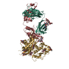 3ngbS S: Starting model for refinement C: citing same article ( |
|---|


-Links
 PDBj
PDBj


- Assembly
| Deposited unit | 
| ||||||||
|---|---|---|---|---|---|---|---|---|---|
| 1 |
| ||||||||
| Unit cell |
|
-Components
| #1: Antibody | Mass: 26690.879 Da / Num. of mol.: 1 Source method: isolated from a genetically manipulated source Source: (gene. exp.) Homo sapiens (human) / Cell (production host): HEK293 / Production host: Homo sapiens (human) |
|---|---|
| #2: Antibody | Mass: 23142.639 Da / Num. of mol.: 1 Source method: isolated from a genetically manipulated source Source: (gene. exp.) Homo sapiens (human) / Cell (production host): HEK293 / Production host: Homo sapiens (human) |
| #3: Water | ChemComp-HOH /  Mass: 18.015 Da / Num. of mol.: 456 / Source method: isolated from a natural source / Formula: H2O Mass: 18.015 Da / Num. of mol.: 456 / Source method: isolated from a natural source / Formula: H2O |
| Has protein modification | Y |
-Experimental details
-Experiment
| Experiment | Method: X-RAY DIFFRACTION / Number of used crystals: 1 |
|---|
- Sample preparation
| Crystal | Density Matthews: 2.31 Å3/Da / Density % sol: 46.67 % |
|---|---|
| Crystal grow | Temperature: 295 K / Method: vapor diffusion, sitting drop / Details: 8% v/v Tacsimate, pH 7.0, 20% w/v PEG3350 |
-Data collection
| Diffraction | Mean temperature: 100 K / Serial crystal experiment: N |
|---|---|
| Diffraction source | Source: SYNCHROTRON / Site: SSRL / Beamline: BL12-2 / Wavelength: 1 Å |
| Detector | Type: DECTRIS EIGER X 16M / Detector: PIXEL / Date: Nov 3, 2022 |
| Radiation | Protocol: SINGLE WAVELENGTH / Monochromatic (M) / Laue (L): M / Scattering type: x-ray |
| Radiation wavelength | Wavelength: 1 Å / Relative weight: 1 |
| Reflection | Resolution: 1.65→38.7 Å / Num. obs: 52273 / % possible obs: 97 % / Redundancy: 6.6 % / CC1/2: 0.99 / Rmerge(I) obs: 0.12 / Rpim(I) all: 0.08 / Net I/σ(I): 7.7 |
| Reflection shell | Resolution: 1.65→1.68 Å / Redundancy: 6.1 % / Rmerge(I) obs: 0.71 / Mean I/σ(I) obs: 1.8 / Num. unique obs: 2575 / CC1/2: 0.82 / Rpim(I) all: 0.45 / % possible all: 96 |
- Processing
| Software |
| ||||||||||||||||
|---|---|---|---|---|---|---|---|---|---|---|---|---|---|---|---|---|---|
| Refinement | Method to determine structure: MOLECULAR REPLACEMENT Starting model: PDB entry 3NGB Resolution: 1.75→38.7 Å / Cross valid method: FREE R-VALUE
| ||||||||||||||||
| Refinement step | Cycle: LAST / Resolution: 1.75→38.7 Å
|
