 Movie
Movie Controller

[English] 日本語
 Yorodumi
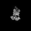
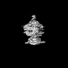
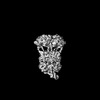
- PDB-8ugb: Cryo-EM structure of bovine phosphodiesterase 6 bound to udenafil -
+ Open data

- Basic information
| Entry | Database: PDB / ID: 8ugb | ||||||
|---|---|---|---|---|---|---|---|
| Title | Cryo-EM structure of bovine phosphodiesterase 6 bound to udenafil | ||||||
 Components Components |
| ||||||
 Keywords Keywords | SIGNALING PROTEIN/INHIBITOR / phosphodiesterase / GPCR effector enzyme / SIGNALING PROTEIN / inhibitor / SIGNALING PROTEIN-INHIBITOR complex | ||||||
| Function / homology |  Function and homology information Function and homology information3',5'-cyclic-GMP phosphodiesterase / Inactivation, recovery and regulation of the phototransduction cascade / Activation of the phototransduction cascade / positive regulation of G protein-coupled receptor signaling pathway / Ca2+ pathway / photoreceptor outer segment membrane / entrainment of circadian clock by photoperiod / retina development in camera-type eye / cGMP binding / 3',5'-cyclic-GMP phosphodiesterase activity ...3',5'-cyclic-GMP phosphodiesterase / Inactivation, recovery and regulation of the phototransduction cascade / Activation of the phototransduction cascade / positive regulation of G protein-coupled receptor signaling pathway / Ca2+ pathway / photoreceptor outer segment membrane / entrainment of circadian clock by photoperiod / retina development in camera-type eye / cGMP binding / 3',5'-cyclic-GMP phosphodiesterase activity / 3',5'-cyclic-AMP phosphodiesterase activity / photoreceptor outer segment / positive regulation of epidermal growth factor receptor signaling pathway / negative regulation of cAMP/PKA signal transduction / visual perception / enzyme inhibitor activity / photoreceptor disc membrane / molecular adaptor activity / signal transduction / zinc ion binding / metal ion binding Similarity search - Function | ||||||
| Biological species |  | ||||||
| Method | ELECTRON MICROSCOPY / single particle reconstruction / cryo EM / Resolution: 3 Å | ||||||
 Authors Authors | Aplin, C. / Cerione, R.A. | ||||||
| Funding support |  United States, 1items United States, 1items
| ||||||
 Citation Citation | Journal: J Biol Chem / Year: 2024 Title: Probing the mechanism by which the retinal G protein transducin activates its biological effector PDE6. Authors: Cody Aplin / Richard A Cerione / Abstract: Phototransduction in retinal rods occurs when the G protein-coupled photoreceptor rhodopsin triggers the activation of phosphodiesterase 6 (PDE6) by GTP-bound alpha subunits of the G protein ...Phototransduction in retinal rods occurs when the G protein-coupled photoreceptor rhodopsin triggers the activation of phosphodiesterase 6 (PDE6) by GTP-bound alpha subunits of the G protein transducin (Gα). Recently, we presented a cryo-EM structure for a complex between two GTP-bound recombinant Gα subunits and native PDE6, that included a bivalent antibody bound to the C-terminal ends of Gα and the inhibitor vardenafil occupying the active sites on the PDEα and PDEβ subunits. We proposed Gα-activated PDE6 by inducing a striking reorientation of the PDEγ subunits away from the catalytic sites. However, questions remained including whether in the absence of the antibody Gα binds to PDE6 in a similar manner as observed when the antibody is present, does Gα activate PDE6 by enabling the substrate cGMP to access the catalytic sites, and how does the lipid membrane enhance PDE6 activation? Here, we demonstrate that 2:1 Gα-PDE6 complexes form with either recombinant or retinal Gα in the absence of the Gα antibody. We show that Gα binding is not necessary for cGMP nor competitive inhibitors to access the active sites; instead, occupancy of the substrate binding sites enables Gα to bind and reposition the PDE6γ subunits to promote catalytic activity. Moreover, we demonstrate by reconstituting Gα-stimulated PDE6 activity in lipid bilayer nanodiscs that the membrane-induced enhancement results from an increase in the apparent binding affinity of Gα for PDE6. These findings provide new insights into how the retinal G protein stimulates rapid catalytic turnover by PDE6 required for dim light vision. | ||||||
| History |
|
- Structure visualization
| Structure viewer | Molecule: MolmilJmol/JSmol |
|---|
- Downloads & links
-Download
| PDBx/mmCIF format | 8ugb.cif.gz | 431.6 KB | Display | PDBx/mmCIF format |
|---|---|---|---|---|
| PDB format | pdb8ugb.ent.gz | 279.2 KB | Display | PDB format |
| PDBx/mmJSON format | 8ugb.json.gz | Tree view | PDBx/mmJSON format | |
| Others |  Other downloads Other downloads |
-Validation report
| Arichive directory | https://data.pdbj.org/pub/pdb/validation_reports/ug/8ugbftp://data.pdbj.org/pub/pdb/validation_reports/ug/8ugb | HTTPS FTP |
|---|
-Related structure data
| Related structure data |  42220MC 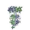 8ufiC 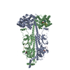 8ugsC 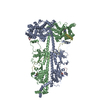 8ulgC M: map data used to model this data C: citing same article ( |
|---|---|
| Similar structure data |


-Links
 PDBj
PDBj


- Assembly
| Deposited unit | 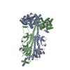
|
|---|---|
| 1 |
|
-Components
-Rod cGMP-specific 3',5'-cyclic phosphodiesterase subunit ... , 2 types, 2 molecules AB
| #1: Protein | Mass: 99461.789 Da / Num. of mol.: 1 / Source method: isolated from a natural source / Source: (natural) References: UniProt: P11541, 3',5'-cyclic-GMP phosphodiesterase |
|---|---|
| #2: Protein | Mass: 98449.648 Da / Num. of mol.: 1 / Source method: isolated from a natural source / Source: (natural) References: UniProt: P23439, 3',5'-cyclic-GMP phosphodiesterase |
-Protein , 1 types, 2 molecules CD
| #3: Protein | Mass: 9684.229 Da / Num. of mol.: 2 / Source method: isolated from a natural source / Source: (natural) References: UniProt: P04972, 3',5'-cyclic-GMP phosphodiesterase |
|---|
-Non-polymers , 4 types, 8 molecules 


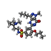
| #4: Chemical |  Mass: 345.205 Da / Num. of mol.: 2 / Source method: obtained synthetically / Formula: C10H12N5O7P / Feature type: SUBJECT OF INVESTIGATION Mass: 345.205 Da / Num. of mol.: 2 / Source method: obtained synthetically / Formula: C10H12N5O7P / Feature type: SUBJECT OF INVESTIGATION#5: Chemical |  Mass: 65.409 Da / Num. of mol.: 2 / Source method: obtained synthetically / Formula: Zn / Feature type: SUBJECT OF INVESTIGATION Mass: 65.409 Da / Num. of mol.: 2 / Source method: obtained synthetically / Formula: Zn / Feature type: SUBJECT OF INVESTIGATION#6: Chemical |  Mass: 24.305 Da / Num. of mol.: 2 / Source method: obtained synthetically / Formula: Mg / Feature type: SUBJECT OF INVESTIGATION Mass: 24.305 Da / Num. of mol.: 2 / Source method: obtained synthetically / Formula: Mg / Feature type: SUBJECT OF INVESTIGATION#7: Chemical |  Mass: 516.656 Da / Num. of mol.: 2 / Source method: obtained synthetically / Formula: C25H36N6O4S / Feature type: SUBJECT OF INVESTIGATION / Comment: inhibitor*YM Mass: 516.656 Da / Num. of mol.: 2 / Source method: obtained synthetically / Formula: C25H36N6O4S / Feature type: SUBJECT OF INVESTIGATION / Comment: inhibitor*YM |
|---|
-Details
| Has ligand of interest | Y |
|---|
-Experimental details
-Experiment
| Experiment | Method: ELECTRON MICROSCOPY |
|---|---|
| EM experiment | Aggregation state: PARTICLE / 3D reconstruction method: single particle reconstruction |
- Sample preparation
| Component | Name: Bovine rod phosphodiesterase 6 bound to udenafil / Type: COMPLEX / Entity ID: #3, #1-#2 / Source: NATURAL |
|---|---|
| Molecular weight | Experimental value: NO |
| Source (natural) | Organism: |
| Buffer solution | pH: 8 |
| Specimen | Embedding applied: NO / Shadowing applied: NO / Staining applied: NO / Vitrification applied: YES |
| Vitrification | Cryogen name: ETHANE |
- Electron microscopy imaging
| Experimental equipment |  Model: Talos Arctica / Image courtesy: FEI Company |
|---|---|
| Microscopy | Model: FEI TALOS ARCTICA |
| Electron gun | Electron source:  FIELD EMISSION GUN / Accelerating voltage: 200 kV / Illumination mode: FLOOD BEAM FIELD EMISSION GUN / Accelerating voltage: 200 kV / Illumination mode: FLOOD BEAM |
| Electron lens | Mode: BRIGHT FIELD / Nominal magnification: 63000 X / Nominal defocus max: 2000 nm / Nominal defocus min: 1400 nm |
| Image recording | Electron dose: 50 e/Å2 / Film or detector model: GATAN K3 (6k x 4k) |
- Processing
| CTF correction | Type: PHASE FLIPPING AND AMPLITUDE CORRECTION | ||||||||||||||||||||||||
|---|---|---|---|---|---|---|---|---|---|---|---|---|---|---|---|---|---|---|---|---|---|---|---|---|---|
| 3D reconstruction | Resolution: 3 Å / Resolution method: FSC 0.143 CUT-OFF / Num. of particles: 2050380 / Symmetry type: POINT | ||||||||||||||||||||||||
| Refinement | Cross valid method: NONE Stereochemistry target values: GeoStd + Monomer Library + CDL v1.2 | ||||||||||||||||||||||||
| Displacement parameters | Biso mean: 95.37 Å2 | ||||||||||||||||||||||||
| Refine LS restraints |
|
