 Movie
Movie Controller


+ Open data

- Basic information
| Entry | Database: PDB / ID: 8ufa | |||||||||||||||||||||||||||||||||||||||||||||||||||||||||||||||
|---|---|---|---|---|---|---|---|---|---|---|---|---|---|---|---|---|---|---|---|---|---|---|---|---|---|---|---|---|---|---|---|---|---|---|---|---|---|---|---|---|---|---|---|---|---|---|---|---|---|---|---|---|---|---|---|---|---|---|---|---|---|---|---|---|
| Title | Eastern equine encephalitis virus (PE-6) VLP (asymmetric unit) | |||||||||||||||||||||||||||||||||||||||||||||||||||||||||||||||
 Components Components |
| |||||||||||||||||||||||||||||||||||||||||||||||||||||||||||||||
 Keywords Keywords | VIRAL PROTEIN / virus / Structural Genomics / Center for Structural Biology of Infectious Diseases / CSBID | |||||||||||||||||||||||||||||||||||||||||||||||||||||||||||||||
| Function / homology |  Function and homology information Function and homology informationtogavirin / T=4 icosahedral viral capsid / host cell endoplasmic reticulum / channel activity / monoatomic ion transmembrane transport / symbiont-mediated suppression of host toll-like receptor signaling pathway / host cell cytoplasm / host cell Golgi apparatus / entry receptor-mediated virion attachment to host cell / symbiont-mediated suppression of host gene expression ...togavirin / T=4 icosahedral viral capsid / host cell endoplasmic reticulum / channel activity / monoatomic ion transmembrane transport / symbiont-mediated suppression of host toll-like receptor signaling pathway / host cell cytoplasm / host cell Golgi apparatus / entry receptor-mediated virion attachment to host cell / symbiont-mediated suppression of host gene expression / serine-type endopeptidase activity / fusion of virus membrane with host endosome membrane / symbiont entry into host cell / virion attachment to host cell / host cell nucleus / host cell plasma membrane / virion membrane / structural molecule activity / proteolysis / RNA binding Similarity search - Function | |||||||||||||||||||||||||||||||||||||||||||||||||||||||||||||||
| Biological species |  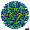 Eastern equine encephalitis virus Eastern equine encephalitis virus | |||||||||||||||||||||||||||||||||||||||||||||||||||||||||||||||
| Method | ELECTRON MICROSCOPY / single particle reconstruction / cryo EM / Resolution: 2.86 Å | |||||||||||||||||||||||||||||||||||||||||||||||||||||||||||||||
 Authors Authors | Adams, L.J. / Fremont, D.H. / Center for Structural Biology of Infectious Diseases (CSBID) | |||||||||||||||||||||||||||||||||||||||||||||||||||||||||||||||
| Funding support |  United States, 1items United States, 1items
| |||||||||||||||||||||||||||||||||||||||||||||||||||||||||||||||
 Citation Citation | Journal: Cell / Year: 2024 Title: Structural and functional basis of VLDLR usage by Eastern equine encephalitis virus. Authors: Lucas J Adams / Saravanan Raju / Hongming Ma / Theron Gilliland / Douglas S Reed / William B Klimstra / Daved H Fremont / Michael S Diamond / Abstract: The very-low-density lipoprotein receptor (VLDLR) comprises eight LDLR type A (LA) domains and supports entry of distantly related alphaviruses, including Eastern equine encephalitis virus (EEEV) and ...The very-low-density lipoprotein receptor (VLDLR) comprises eight LDLR type A (LA) domains and supports entry of distantly related alphaviruses, including Eastern equine encephalitis virus (EEEV) and Semliki Forest virus (SFV). Here, by resolving multiple cryo-electron microscopy structures of EEEV-VLDLR complexes and performing mutagenesis and functional studies, we show that EEEV uses multiple sites (E1/E2 cleft and E2 A domain) to engage more than one LA domain simultaneously. However, no single LA domain is necessary or sufficient to support efficient EEEV infection. Whereas all EEEV strains show conservation of two VLDLR-binding sites, the EEEV PE-6 strain and a few other EEE complex members feature a single amino acid substitution that enables binding of LA domains to an additional site on the E2 B domain. These structural and functional analyses informed the design of a minimal VLDLR decoy receptor that neutralizes EEEV infection and protects mice from lethal challenge. | |||||||||||||||||||||||||||||||||||||||||||||||||||||||||||||||
| History |
|
- Structure visualization
| Structure viewer | Molecule: MolmilJmol/JSmol |
|---|
- Downloads & links
-Download
| PDBx/mmCIF format | 8ufa.cif.gz | 668.4 KB | Display | PDBx/mmCIF format |
|---|---|---|---|---|
| PDB format | pdb8ufa.ent.gz | 531.5 KB | Display | PDB format |
| PDBx/mmJSON format | 8ufa.json.gz | Tree view | PDBx/mmJSON format | |
| Others |  Other downloads Other downloads |
-Validation report
| Arichive directory | https://data.pdbj.org/pub/pdb/validation_reports/uf/8ufaftp://data.pdbj.org/pub/pdb/validation_reports/uf/8ufa | HTTPS FTP |
|---|
-Related structure data
| Related structure data |  42188MC  8ufbC  8ufcC M: map data used to model this data C: citing same article ( |
|---|---|
| Similar structure data |


-Links
 PDBj
PDBj


- Assembly
| Deposited unit | 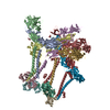
|
|---|---|
| 1 |
|
-Components
| #1: Protein | Mass: 47935.180 Da / Num. of mol.: 4 / Fragment: UNP residues 802-1242 Source method: isolated from a genetically manipulated source Source: (gene. exp.) Eastern equine encephalitis virus / Strain: PE-6 / Production host:  Homo sapiens (human) / References: UniProt: Q88678 Homo sapiens (human) / References: UniProt: Q88678#2: Protein | Mass: 46378.191 Da / Num. of mol.: 4 / Fragment: UNP residues 325-738 Source method: isolated from a genetically manipulated source Source: (gene. exp.) Eastern equine encephalitis virus / Strain: PE-6 / Production host: Homo sapiens (human) / References: UniProt: Q88678#3: Protein | Mass: 16540.656 Da / Num. of mol.: 4 / Fragment: UNP residues 111-261 Source method: isolated from a genetically manipulated source Source: (gene. exp.) Eastern equine encephalitis virus / Strain: PE-6 / Gene: SP / Production host: Homo sapiens (human) / References: UniProt: W8S146#4: Sugar | ChemComp-NAG /   Type: D-saccharide, beta linking / Mass: 221.208 Da / Num. of mol.: 8 / Source method: obtained synthetically / Formula: C8H15NO6 Type: D-saccharide, beta linking / Mass: 221.208 Da / Num. of mol.: 8 / Source method: obtained synthetically / Formula: C8H15NO6Has ligand of interest | N | Has protein modification | Y | |
|---|
-Experimental details
-Experiment
| Experiment | Method: ELECTRON MICROSCOPY |
|---|---|
| EM experiment | Aggregation state: PARTICLE / 3D reconstruction method: single particle reconstruction |
- Sample preparation
| Component | Name: Eastern equine encephalitis virus / Type: VIRUS / Entity ID: #1-#3 / Source: RECOMBINANT |
|---|---|
| Source (natural) | Organism: Eastern equine encephalitis virus / Strain: PE-6 |
| Source (recombinant) | Organism: Homo sapiens (human) |
| Details of virus | Empty: YES / Enveloped: YES / Isolate: STRAIN / Type: VIRUS-LIKE PARTICLE |
| Buffer solution | pH: 7.4 |
| Specimen | Embedding applied: NO / Shadowing applied: NO / Staining applied: NO / Vitrification applied: YES |
| Vitrification | Cryogen name: ETHANE |
- Electron microscopy imaging
| Experimental equipment |  Model: Titan Krios / Image courtesy: FEI Company |
|---|---|
| Microscopy | Model: FEI TITAN KRIOS |
| Electron gun | Electron source:  FIELD EMISSION GUN / Accelerating voltage: 300 kV / Illumination mode: FLOOD BEAM FIELD EMISSION GUN / Accelerating voltage: 300 kV / Illumination mode: FLOOD BEAM |
| Electron lens | Mode: BRIGHT FIELD / Nominal defocus max: 2200 nm / Nominal defocus min: 700 nm |
| Image recording | Electron dose: 37.93 e/Å2 / Film or detector model: FEI FALCON IV (4k x 4k) |
- Processing
| EM software | Name: PHENIX / Category: model refinement | ||||||||||||||||||||||||
|---|---|---|---|---|---|---|---|---|---|---|---|---|---|---|---|---|---|---|---|---|---|---|---|---|---|
| CTF correction | Type: PHASE FLIPPING AND AMPLITUDE CORRECTION | ||||||||||||||||||||||||
| 3D reconstruction | Resolution: 2.86 Å / Resolution method: FSC 0.143 CUT-OFF / Num. of particles: 1038056 / Symmetry type: POINT | ||||||||||||||||||||||||
| Refine LS restraints |
|
