Movie
Movie Controller

[English] 日本語
 Yorodumi
- PDB-8ubv: Cryo-EM structure of dimeric FBXL17-BACH1BTB E3 ubiquitin ligase ... -
+ Open data

- Basic information
| Entry | Database: PDB / ID: 8ubv | ||||||
|---|---|---|---|---|---|---|---|
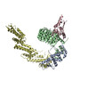
| Title | Cryo-EM structure of dimeric FBXL17-BACH1BTB E3 ubiquitin ligase complex | ||||||
 Components Components |
| ||||||
 Keywords Keywords | LIGASE / FBXL17 / BACH1 / F-box protein / Cullin / E3 ligase | ||||||
| Function / homology |  Function and homology information Function and homology informationRegulation of HMOX1 expression and activity / neural crest cell differentiation / ligand-modulated transcription factor activity / regulation of smoothened signaling pathway / NFE2L2 regulating anti-oxidant/detoxification enzymes / SCF ubiquitin ligase complex / SCF-dependent proteasomal ubiquitin-dependent protein catabolic process / protein quality control for misfolded or incompletely synthesized proteins / Regulation of BACH1 activity / Heme signaling ...Regulation of HMOX1 expression and activity / neural crest cell differentiation / ligand-modulated transcription factor activity / regulation of smoothened signaling pathway / NFE2L2 regulating anti-oxidant/detoxification enzymes / SCF ubiquitin ligase complex / SCF-dependent proteasomal ubiquitin-dependent protein catabolic process / protein quality control for misfolded or incompletely synthesized proteins / Regulation of BACH1 activity / Heme signaling / DNA-binding transcription repressor activity, RNA polymerase II-specific / protein polyubiquitination / nervous system development / DNA-binding transcription activator activity, RNA polymerase II-specific / proteasome-mediated ubiquitin-dependent protein catabolic process / DNA-binding transcription factor activity, RNA polymerase II-specific / RNA polymerase II cis-regulatory region sequence-specific DNA binding / protein ubiquitination / DNA-binding transcription factor activity / DNA repair / heme binding / regulation of transcription by RNA polymerase II / regulation of DNA-templated transcription / chromatin / negative regulation of transcription by RNA polymerase II / DNA-templated transcription / nucleoplasm / nucleus / cytosol / cytoplasm Similarity search - Function | ||||||
| Biological species |  Homo sapiens (human) Homo sapiens (human) | ||||||
| Method | ELECTRON MICROSCOPY / single particle reconstruction / cryo EM / Resolution: 4.1 Å | ||||||
 Authors Authors | Shi, H. / Cao, S. / Zheng, N. | ||||||
| Funding support |  United States, 1items United States, 1items
| ||||||
 Citation Citation | Journal: Cell / Year: 2024 Title: Recognition of BACH1 quaternary structure degrons by two F-box proteins under oxidative stress. Authors: Shiyun Cao / Sheena Faye Garcia / Huigang Shi / Ellie I James / Yuki Kito / Hui Shi / Haibin Mao / Sharon Kaisari / Gergely Rona / Sophia Deng / Hailey V Goldberg / Jackeline Ponce / Beatrix ...Authors: Shiyun Cao / Sheena Faye Garcia / Huigang Shi / Ellie I James / Yuki Kito / Hui Shi / Haibin Mao / Sharon Kaisari / Gergely Rona / Sophia Deng / Hailey V Goldberg / Jackeline Ponce / Beatrix Ueberheide / Luca Lignitto / Miklos Guttman / Michele Pagano / Ning Zheng /   Abstract: Ubiquitin-dependent proteolysis regulates diverse cellular functions with high substrate specificity, which hinges on the ability of ubiquitin E3 ligases to decode the targets' degradation signals, i. ...Ubiquitin-dependent proteolysis regulates diverse cellular functions with high substrate specificity, which hinges on the ability of ubiquitin E3 ligases to decode the targets' degradation signals, i.e., degrons. Here, we show that BACH1, a transcription repressor of antioxidant response genes, features two distinct unconventional degrons encrypted in the quaternary structure of its homodimeric BTB domain. These two degrons are both functionalized by oxidative stress and are deciphered by two complementary E3s. FBXO22 recognizes a degron constructed by the BACH1 BTB domain dimer interface, which is unmasked from transcriptional co-repressors after oxidative stress releases BACH1 from chromatin. When this degron is impaired by oxidation, a second BACH1 degron manifested by its destabilized BTB dimer is probed by a pair of FBXL17 proteins that remodels the substrate into E3-bound monomers for ubiquitination. Our findings highlight the multidimensionality of protein degradation signals and the functional complementarity of different ubiquitin ligases targeting the same substrate. | ||||||
| History |
|
- Structure visualization
| Structure viewer | Molecule: MolmilJmol/JSmol |
|---|
- Downloads & links
-Download
| PDBx/mmCIF format | 8ubv.cif.gz | 159.4 KB | Display | PDBx/mmCIF format |
|---|---|---|---|---|
| PDB format | pdb8ubv.ent.gz | 124.2 KB | Display | PDB format |
| PDBx/mmJSON format | 8ubv.json.gz | Tree view | PDBx/mmJSON format | |
| Others |  Other downloads Other downloads |
-Validation report
| Arichive directory | https://data.pdbj.org/pub/pdb/validation_reports/ub/8ubvftp://data.pdbj.org/pub/pdb/validation_reports/ub/8ubv | HTTPS FTP |
|---|
-Related structure data
| Related structure data |  42106MC  8ua3C  8ua6C  8uahC  8ubtC  8ubuC C: citing same article ( M: map data used to model this data |
|---|---|
| Similar structure data |


-Links
 PDBj
PDBj


- Assembly
| Deposited unit | 
|
|---|---|
| 1 |
|
-Components
| #1: Protein | Mass: 44505.512 Da / Num. of mol.: 2 / Fragment: UNP residues 310-701 Source method: isolated from a genetically manipulated source Source: (gene. exp.) Homo sapiens (human) / Gene: FBXL17 / Cell line (production host): BL21 / Production host:  Trichoplusia ni (cabbage looper) / References: UniProt: Q9UF56 Trichoplusia ni (cabbage looper) / References: UniProt: Q9UF56#2: Protein | Mass: 13839.761 Da / Num. of mol.: 2 / Fragment: BTB domain (UNP residues 7-128) Source method: isolated from a genetically manipulated source Source: (gene. exp.) Homo sapiens (human) / Gene: BACH1 / Production host:  Has protein modification | Y | |
|---|
-Experimental details
-Experiment
| Experiment | Method: ELECTRON MICROSCOPY |
|---|---|
| EM experiment | Aggregation state: PARTICLE / 3D reconstruction method: single particle reconstruction |
- Sample preparation
| Component | Name: The structure of dimeric BFXL17-BACH1BTB / Type: COMPLEX / Entity ID: all / Source: MULTIPLE SOURCES |
|---|---|
| Source (natural) | Organism: Homo sapiens (human) |
| Source (recombinant) | Organism: Trichoplusia ni (cabbage looper) |
| Buffer solution | pH: 7.5 |
| Specimen | Embedding applied: NO / Shadowing applied: NO / Staining applied: NO / Vitrification applied: YES |
| Vitrification | Instrument: FEI VITROBOT MARK IV / Cryogen name: ETHANE |
- Electron microscopy imaging
| Experimental equipment |  Model: Titan Krios / Image courtesy: FEI Company |
|---|---|
| Microscopy | Model: TFS KRIOS |
| Electron gun | Electron source:  FIELD EMISSION GUN / Accelerating voltage: 300 kV / Illumination mode: FLOOD BEAM FIELD EMISSION GUN / Accelerating voltage: 300 kV / Illumination mode: FLOOD BEAM |
| Electron lens | Mode: BRIGHT FIELD / Nominal defocus max: 25000 nm / Nominal defocus min: 5000 nm / Alignment procedure: COMA FREE |
| Specimen holder | Cryogen: NITROGEN / Specimen holder model: FEI TITAN KRIOS AUTOGRID HOLDER |
| Image recording | Electron dose: 60 e/Å2 / Film or detector model: FEI FALCON IV (4k x 4k) |
- Processing
| EM software |
| ||||||||||||||||||||||||
|---|---|---|---|---|---|---|---|---|---|---|---|---|---|---|---|---|---|---|---|---|---|---|---|---|---|
| CTF correction | Type: PHASE FLIPPING AND AMPLITUDE CORRECTION | ||||||||||||||||||||||||
| 3D reconstruction | Resolution: 4.1 Å / Resolution method: FSC 0.143 CUT-OFF / Num. of particles: 71155 / Algorithm: BACK PROJECTION / Symmetry type: POINT | ||||||||||||||||||||||||
| Atomic model building | Protocol: RIGID BODY FIT / Space: REAL | ||||||||||||||||||||||||
| Atomic model building | PDB-ID: 6WCQ Accession code: 6WCQ / Source name: PDB / Type: experimental model |