 Movie
Movie Controller

[English] 日本語
 Yorodumi
- PDB-8u95: The structure of myosin heavy chain from Drosophila melanogaster ... -
+ Open data

- Basic information
| Entry | Database: PDB / ID: 8u95 | |||||||||
|---|---|---|---|---|---|---|---|---|---|---|
| Title | The structure of myosin heavy chain from Drosophila melanogaster flight muscle thick filaments | |||||||||
 Components Components | Myosin heavy chain, isoform U | |||||||||
 Keywords Keywords | STRUCTURAL PROTEIN / Striated muscle / regulatory protein / myosin / flightin / myofilin / stretchin | |||||||||
| Function / homology |  Function and homology information Function and homology informationmyosin ATPase / cellular component assembly / anatomical structure formation involved in morphogenesis / myosin filament / myosin complex / cytoskeletal motor activity / isomerase activity / supramolecular fiber organization / sarcomere / actin filament binding ...myosin ATPase / cellular component assembly / anatomical structure formation involved in morphogenesis / myosin filament / myosin complex / cytoskeletal motor activity / isomerase activity / supramolecular fiber organization / sarcomere / actin filament binding / actin cytoskeleton organization / ATP binding Similarity search - Function | |||||||||
| Biological species |  | |||||||||
| Method | ELECTRON MICROSCOPY / single particle reconstruction / cryo EM / Resolution: 4.7 Å | |||||||||
 Authors Authors | Abbasi Yeganeh, F. / Rastegarpouyani, H. / Li, J. / Taylor, K.A. | |||||||||
| Funding support |  United States, 2items United States, 2items
| |||||||||
 Citation Citation | Journal: Int J Mol Sci / Year: 2023 Title: Structure of the Flight Muscle Myosin Filament at 4.7 Å Resolution Reveals New Details of Non-Myosin Proteins. Authors: Fatemeh Abbasi Yeganeh / Hosna Rastegarpouyani / Jiawei Li / Kenneth A Taylor / Abstract: Striated muscle thick filaments are composed of myosin II and several non-myosin proteins which define the filament length and modify its function. Myosin II has a globular N-terminal motor domain ...Striated muscle thick filaments are composed of myosin II and several non-myosin proteins which define the filament length and modify its function. Myosin II has a globular N-terminal motor domain comprising its catalytic and actin-binding activities and a long α-helical, coiled tail that forms the dense filament backbone. Myosin alone polymerizes into filaments of irregular length, but striated muscle thick filaments have defined lengths that, with thin filaments, define the sarcomere structure. The motor domain structure and function are well understood, but the myosin filament backbone is not. Here we report on the structure of the flight muscle thick filaments from at 4.7 Å resolution, which eliminates previous ambiguities in non-myosin densities. The full proximal S2 region is resolved, as are the connecting densities between the Ig domains of stretchin-klp. The proteins, flightin, and myofilin are resolved in sufficient detail to build an atomic model based on an AlphaFold prediction. Our results suggest a method by which flightin and myofilin cooperate to define the structure of the thick filament and explains a key myosin mutation that affects flightin incorporation. is a genetic model organism for which our results can define strategies for functional testing. | |||||||||
| History |
|
- Structure visualization
| Structure viewer | Molecule: MolmilJmol/JSmol |
|---|
- Downloads & links
-Download
| PDBx/mmCIF format | 8u95.cif.gz | 391.9 KB | Display | PDBx/mmCIF format |
|---|---|---|---|---|
| PDB format | pdb8u95.ent.gz | 303.4 KB | Display | PDB format |
| PDBx/mmJSON format | 8u95.json.gz | Tree view | PDBx/mmJSON format | |
| Others |  Other downloads Other downloads |
-Validation report
| Arichive directory | https://data.pdbj.org/pub/pdb/validation_reports/u9/8u95ftp://data.pdbj.org/pub/pdb/validation_reports/u9/8u95 | HTTPS FTP |
|---|
-Related structure data
| Related structure data | 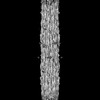 42024MC  8u8hC M: map data used to model this data C: citing same article ( |
|---|---|
| Similar structure data |
-Links
 PDBj
PDBj


- Assembly
| Deposited unit | 
|
|---|---|
| 1 |
|
-Components
| #1: Protein | Mass: 223260.797 Da / Num. of mol.: 2 / Source method: isolated from a natural source / Source: (natural) Has protein modification | N | |
|---|
-Experimental details
-Experiment
| Experiment | Method: ELECTRON MICROSCOPY |
|---|---|
| EM experiment | Aggregation state: FILAMENT / 3D reconstruction method: single particle reconstruction |
- Sample preparation
| Component | Name: Drosophila melanogaster flight muscle thick filament / Type: COMPLEX / Entity ID: all / Source: NATURAL |
|---|---|
| Source (natural) | Organism: |
| Buffer solution | pH: 6.8 |
| Specimen | Embedding applied: NO / Shadowing applied: NO / Staining applied: NO / Vitrification applied: YES |
| Vitrification | Cryogen name: ETHANE |
- Electron microscopy imaging
| Experimental equipment |  Model: Titan Krios / Image courtesy: FEI Company |
|---|---|
| Microscopy | Model: FEI TITAN KRIOS |
| Electron gun | Electron source:  FIELD EMISSION GUN / Accelerating voltage: 300 kV / Illumination mode: FLOOD BEAM FIELD EMISSION GUN / Accelerating voltage: 300 kV / Illumination mode: FLOOD BEAM |
| Electron lens | Mode: BRIGHT FIELD / Nominal defocus max: 2000 nm / Nominal defocus min: 500 nm |
| Image recording | Electron dose: 55 e/Å2 / Film or detector model: GATAN K3 BIOQUANTUM (6k x 4k) |
- Processing
| EM software | Name: PHENIX / Category: model refinement | ||||||||||||||||||||||||
|---|---|---|---|---|---|---|---|---|---|---|---|---|---|---|---|---|---|---|---|---|---|---|---|---|---|
| CTF correction | Type: PHASE FLIPPING AND AMPLITUDE CORRECTION | ||||||||||||||||||||||||
| 3D reconstruction | Resolution: 4.7 Å / Resolution method: OTHER / Num. of particles: 116000 / Symmetry type: POINT | ||||||||||||||||||||||||
| Refine LS restraints |
|
