 Movie
Movie Controller

[English] 日本語
 Yorodumi
- EMDB-42024: CryoEM structure of the Drosophila melanogaster flight muscle myo... -
+ Open data

- Basic information
| Entry | 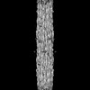 | |||||||||
|---|---|---|---|---|---|---|---|---|---|---|
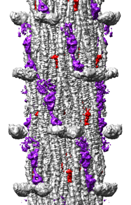
| Title | CryoEM structure of the Drosophila melanogaster flight muscle myosin filament | |||||||||
 Map data Map data | ||||||||||
 Sample Sample |
| |||||||||
 Keywords Keywords | Striated muscle / regulatory protein / myosin / flightin / myofilin / stretchin / STRUCTURAL PROTEIN | |||||||||
| Function / homology |  Function and homology information Function and homology informationmyosin ATPase / oscillatory muscle contraction / cellular component assembly / adult somatic muscle development / striated muscle myosin thick filament assembly / striated muscle myosin thick filament / anatomical structure formation involved in morphogenesis / structural molecule activity conferring elasticity / A band / myosin filament ...myosin ATPase / oscillatory muscle contraction / cellular component assembly / adult somatic muscle development / striated muscle myosin thick filament assembly / striated muscle myosin thick filament / anatomical structure formation involved in morphogenesis / structural molecule activity conferring elasticity / A band / myosin filament / myosin complex / structural constituent of muscle / sarcomere organization / cytoskeletal motor activity / isomerase activity / supramolecular fiber organization / sarcomere / actin filament binding / actin cytoskeleton organization / ATP binding Similarity search - Function | |||||||||
| Biological species |  | |||||||||
| Method | single particle reconstruction / cryo EM / Resolution: 4.7 Å | |||||||||
 Authors Authors | Abbasi Yeganeh F / Rastegarpouyani H / Li J / Taylor KA | |||||||||
| Funding support |  United States, 2 items United States, 2 items
| |||||||||
 Citation Citation | Journal: Int J Mol Sci / Year: 2023 Title: Structure of the Flight Muscle Myosin Filament at 4.7 Å Resolution Reveals New Details of Non-Myosin Proteins. Authors: Fatemeh Abbasi Yeganeh / Hosna Rastegarpouyani / Jiawei Li / Kenneth A Taylor / Abstract: Striated muscle thick filaments are composed of myosin II and several non-myosin proteins which define the filament length and modify its function. Myosin II has a globular N-terminal motor domain ...Striated muscle thick filaments are composed of myosin II and several non-myosin proteins which define the filament length and modify its function. Myosin II has a globular N-terminal motor domain comprising its catalytic and actin-binding activities and a long α-helical, coiled tail that forms the dense filament backbone. Myosin alone polymerizes into filaments of irregular length, but striated muscle thick filaments have defined lengths that, with thin filaments, define the sarcomere structure. The motor domain structure and function are well understood, but the myosin filament backbone is not. Here we report on the structure of the flight muscle thick filaments from at 4.7 Å resolution, which eliminates previous ambiguities in non-myosin densities. The full proximal S2 region is resolved, as are the connecting densities between the Ig domains of stretchin-klp. The proteins, flightin, and myofilin are resolved in sufficient detail to build an atomic model based on an AlphaFold prediction. Our results suggest a method by which flightin and myofilin cooperate to define the structure of the thick filament and explains a key myosin mutation that affects flightin incorporation. is a genetic model organism for which our results can define strategies for functional testing. | |||||||||
| History |
|
- Structure visualization
| Supplemental images |
|---|
- Downloads & links
-EMDB archive
| Map data | emd_42024.map.gz | 1.6 GB | EMDB map data format | |
|---|---|---|---|---|
| Header (meta data) | emd-42024-v30.xmlemd-42024.xml | 19.8 KB 19.8 KB | Display Display | EMDB header |
| Images |  emd_42024.png emd_42024.png | 149.3 KB | ||
| Filedesc metadata | emd-42024.cif.gz | 6.1 KB | ||
| Others | emd_42024_additional_1.map.gzemd_42024_additional_2.map.gzemd_42024_half_map_1.map.gzemd_42024_half_map_2.map.gz | 609.4 KB 31.8 KB 1.2 GB 1.2 GB | ||
| Archive directory |  http://ftp.pdbj.org/pub/emdb/structures/EMD-42024ftp://ftp.pdbj.org/pub/emdb/structures/EMD-42024 http://ftp.pdbj.org/pub/emdb/structures/EMD-42024ftp://ftp.pdbj.org/pub/emdb/structures/EMD-42024 | HTTPS FTP |
-Related structure data
| Related structure data |  8u8hMC  8u95MC M: atomic model generated by this map C: citing same article ( |
|---|---|
| Similar structure data |
-Links
| EMDB pages | EMDB (EBI/PDBe) / EMDataResource |
|---|---|
| Related items in Molecule of the Month |


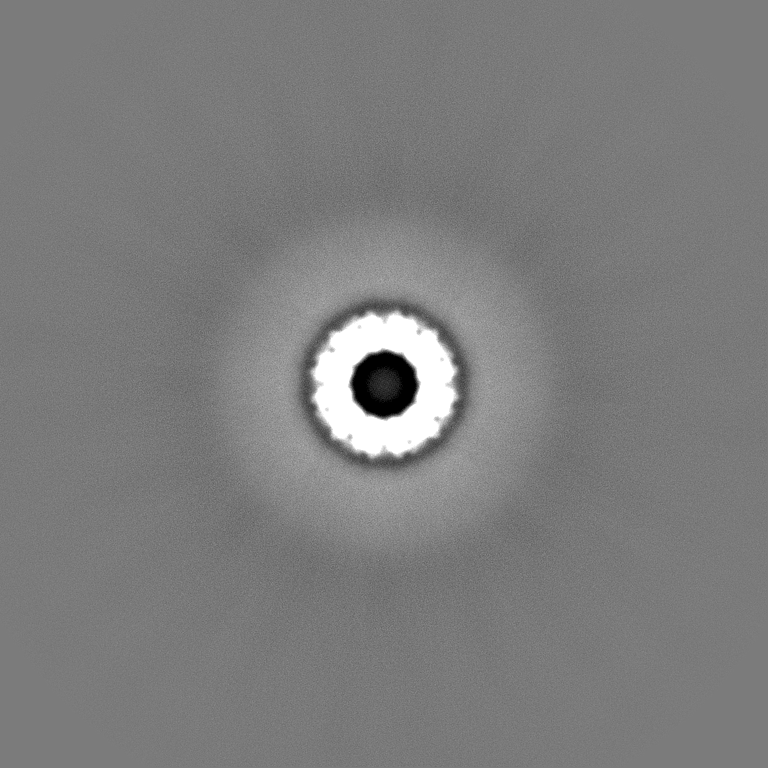
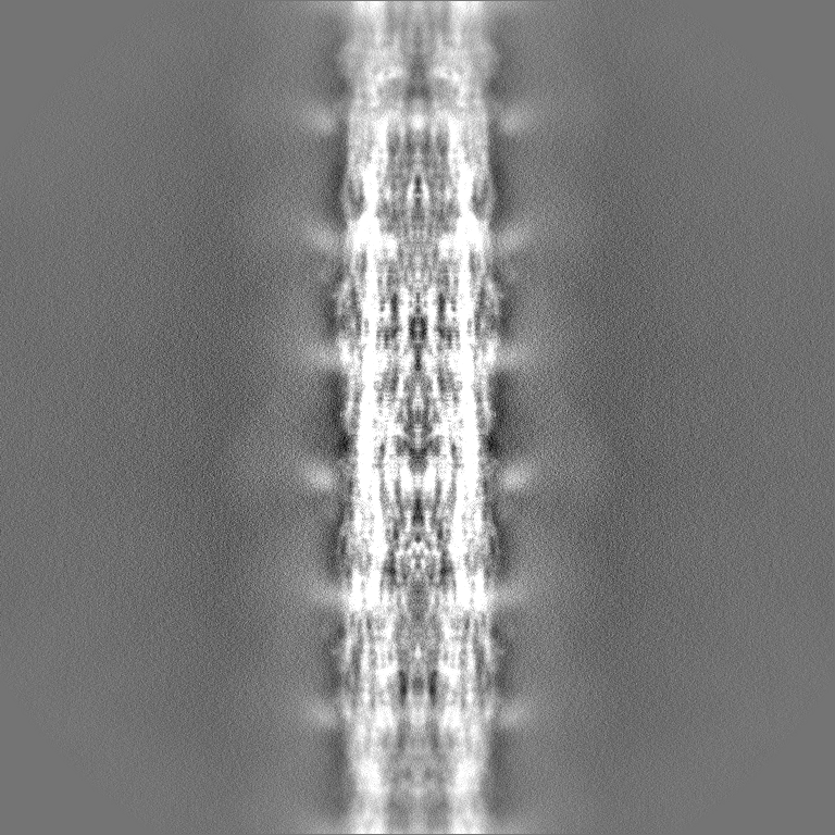
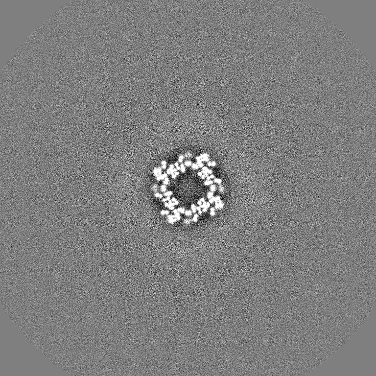
-Map
| File | Download / File: emd_42024.map.gz / Format: CCP4 / Size: 1.7 GB / Type: IMAGE STORED AS FLOATING POINT NUMBER (4 BYTES) | ||||||||||||||||||||||||||||||||||||
|---|---|---|---|---|---|---|---|---|---|---|---|---|---|---|---|---|---|---|---|---|---|---|---|---|---|---|---|---|---|---|---|---|---|---|---|---|---|
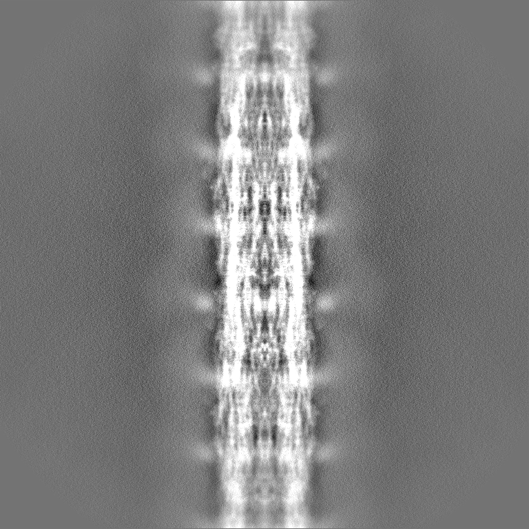
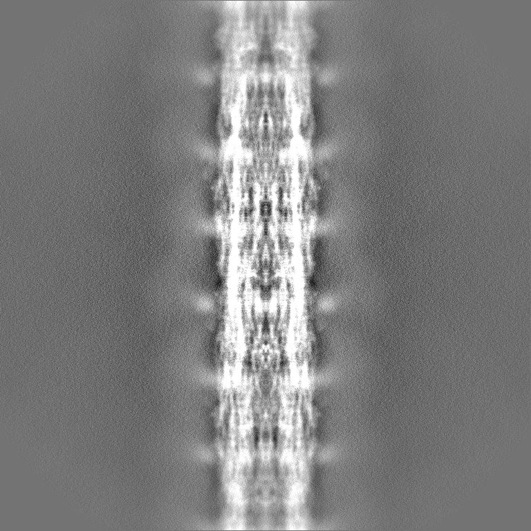
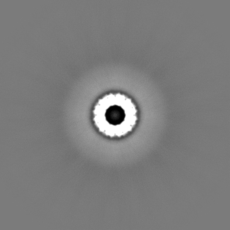
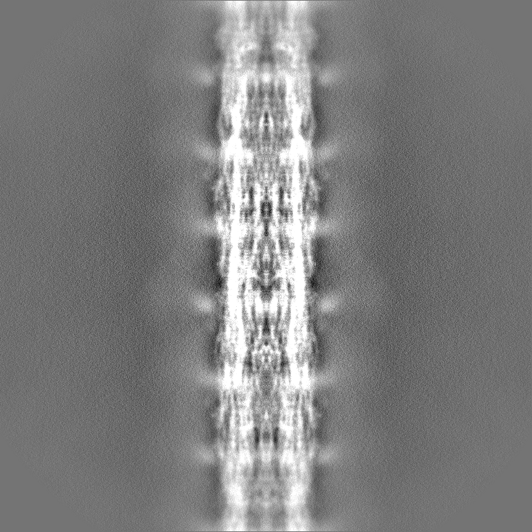
| Projections & slices | Image control
Images are generated by Spider. | ||||||||||||||||||||||||||||||||||||
| Voxel size | X=Y=Z: 1.33 Å | ||||||||||||||||||||||||||||||||||||
| Density |
| ||||||||||||||||||||||||||||||||||||
| Symmetry | Space group: 1 | ||||||||||||||||||||||||||||||||||||
| Details | EMDB XML:
|
 Z (Sec.)
 Y (Row.)
 X (Col.)


-Supplemental data
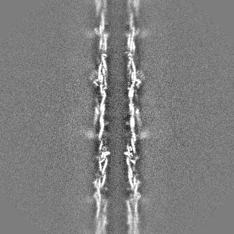
-Additional map: #1
| File | emd_42024_additional_1.map | ||||||||||||
|---|---|---|---|---|---|---|---|---|---|---|---|---|---|
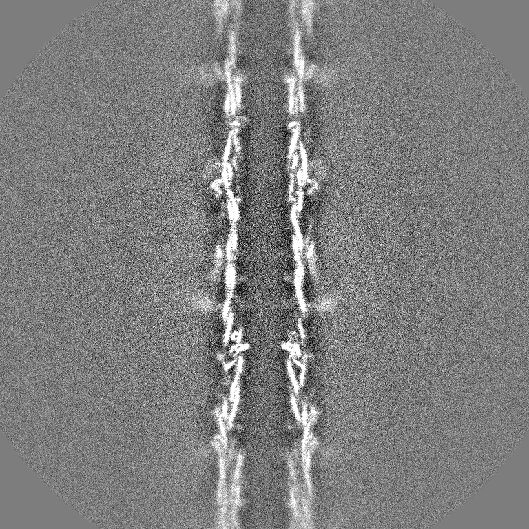
| Projections & Slices |
| ||||||||||||
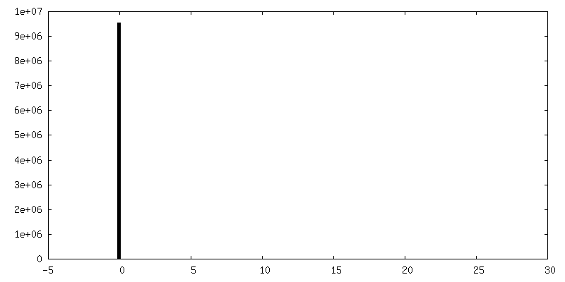
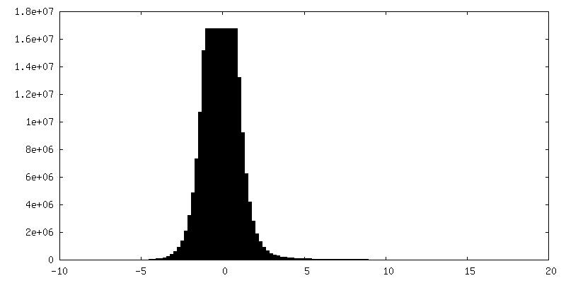
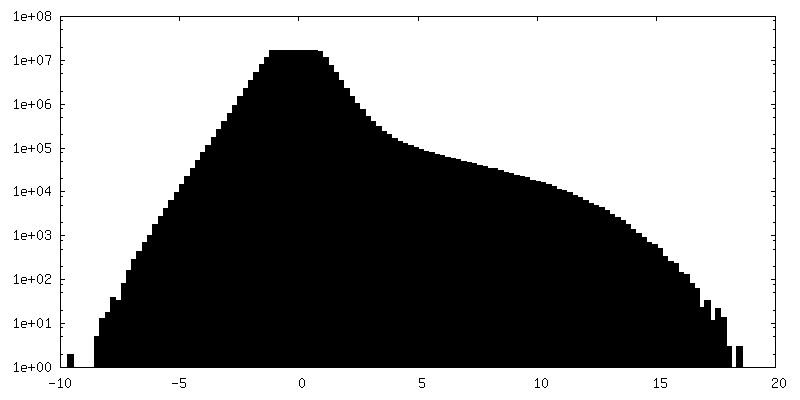
| Density Histograms |


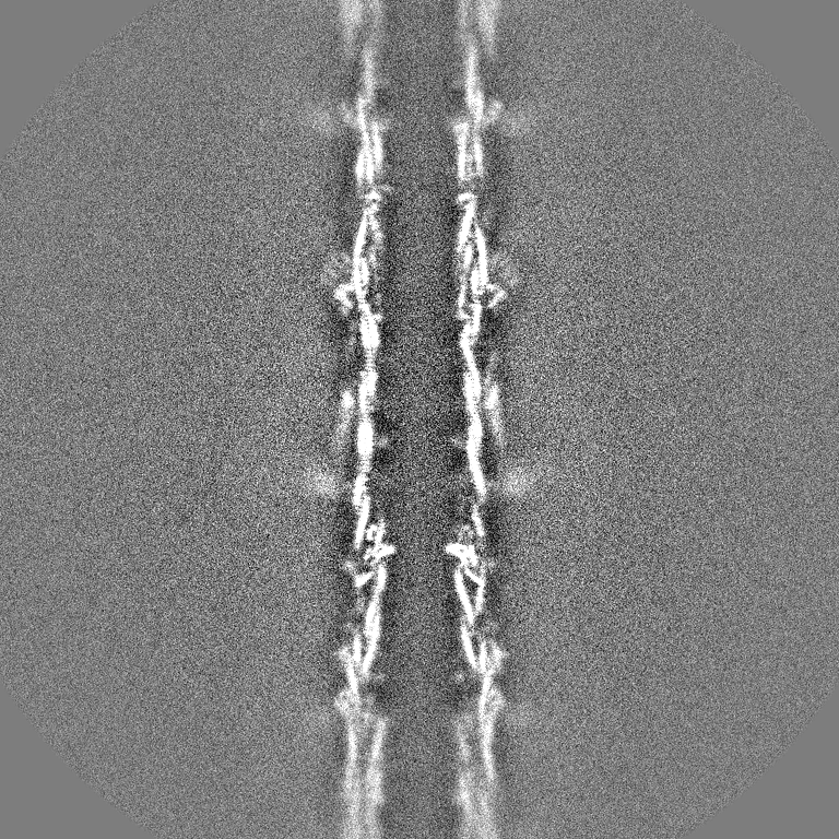
-Additional map: #2
| File | emd_42024_additional_2.map | ||||||||||||
|---|---|---|---|---|---|---|---|---|---|---|---|---|---|
| Projections & Slices |
| ||||||||||||
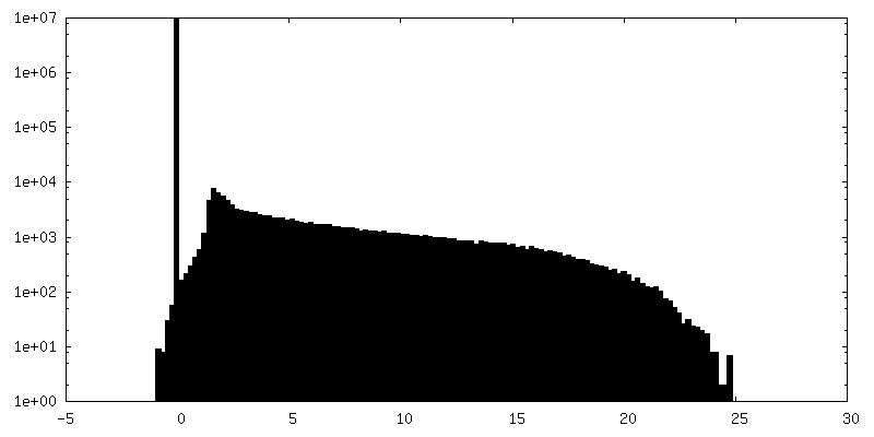
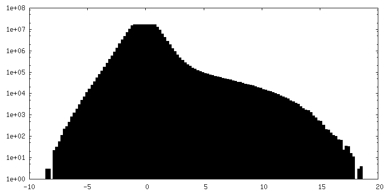
| Density Histograms |


-Half map: #1
| File | emd_42024_half_map_1.map | ||||||||||||
|---|---|---|---|---|---|---|---|---|---|---|---|---|---|
| Projections & Slices |
| ||||||||||||
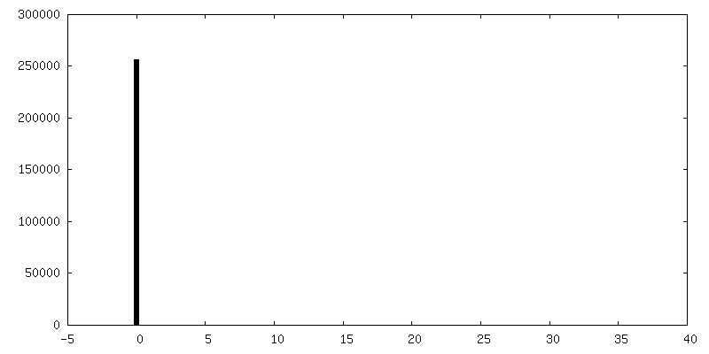
| Density Histograms |


-Half map: #2
| File | emd_42024_half_map_2.map | ||||||||||||
|---|---|---|---|---|---|---|---|---|---|---|---|---|---|
| Projections & Slices |
| ||||||||||||
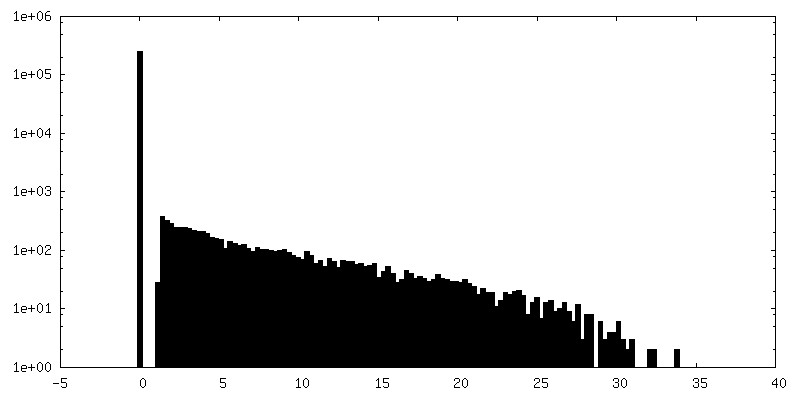
| Density Histograms |

- Sample components
-Entire : Drosophila melanogaster flight muscle thick filament
| Entire | Name: Drosophila melanogaster flight muscle thick filament |
|---|---|
| Components |
|
-Supramolecule #1: Drosophila melanogaster flight muscle thick filament
| Supramolecule | Name: Drosophila melanogaster flight muscle thick filament / type: complex / ID: 1 / Parent: 0 / Macromolecule list: all |
|---|---|
| Source (natural) | Organism: |
-Macromolecule #1: Flightin
| Macromolecule | Name: Flightin / type: protein_or_peptide / ID: 1 / Enantiomer: LEVO |
|---|---|
| Source (natural) | Organism: |
| Sequence | String: MADEEDPWGF DDGGEEEKAA STQAGTPAPP SKAPSVASDH KADSVVAGTP ANEEAAPEEV EEIKAPPPP PEDDGYRKPV QLYRHWVRPK FLQYKYMYNY RTNYYDDVID YIDKKQTGVA R EIPRPQTW AERVLRTRNI SGSDIDSYAP AKRDKQLIQT LAASIRTYNY HTKAYINQRY AS VL |
-Macromolecule #2: Myosin
| Macromolecule | Name: Myosin / type: protein_or_peptide / ID: 2 / Enantiomer: LEVO |
|---|---|
| Source (natural) | Organism: |
| Sequence | String: MPKPVANQED EDPTPYLFVS LEQRRIDQSK PYDSKKSCWI PDEKEGYLLG EIKATKGDIV SVGLQGGEV RDIKSEKVEK VNPPKFEKIE DMADMTVLNT PCVLHNLRQR YYAKLIYTYS G LFCVAINP YKRYPVYTNR CAKMYRGKRR NEVPPHIFAI SDGAYVDMLT ...String: MPKPVANQED EDPTPYLFVS LEQRRIDQSK PYDSKKSCWI PDEKEGYLLG EIKATKGDIV SVGLQGGEV RDIKSEKVEK VNPPKFEKIE DMADMTVLNT PCVLHNLRQR YYAKLIYTYS G LFCVAINP YKRYPVYTNR CAKMYRGKRR NEVPPHIFAI SDGAYVDMLT NHVNQSMLIT GE SGAGKTE NTKKVIAYFA TVGASKKTDE AAKSKGSLED QVVQTNPVLE AFGNAKTVRN DNS SRFGKF IRIHFGPTGK LAGADIETYL LEKARVISQQ SLERSYHIFY QIMSGSVPGV KDIC LLTDN IYDYHIVSQG KVTVASIDDA EEFSLTDQAF DILGFTKQEK EDVYRITAAV MHMGG MKFK QRGREEQAEQ DGEEEGGRVS KLFGCDTAEL YKNLLKPRIK VGNEFVTQGR NVQQVT NSI GALCKGVFDR LFKWLVKKCN ETLDTQQKRQ HFIGVLDIAG FEIFEYNGFE QLCINFT NE KLQQFFNHIM FVMEQEEYKK EGINWDFIDF GMDLLACIDL IEKPMGILSI LEEESMFP K ATDQTFSEKL TNTHLGKSAP FQKPKPPKPG QQAAHFAIAH YAGCVSYNIT GWLEKNKDP LNDTVVDQFK KSQNKLLIEI FADHAGQSGG GEQAKGGRGK KGGGFATVSS AYKEQLNSLM TTLRSTQPH FVRCIIPNEM KQPGVVDAHL VMHQLTCNGV LEGIRICRKG FPNRMMYPDF K MRYQILNP RGIKDLDCPK KASKVLIEST ELNEDLYRLG HTKVFFRAGV LGQMEEFRDE RL GKIMSWM QAWARGYLSR KGFKKLQEQR VALKVVQRNL RKYLQLRTWP WYKLWQKVKP LLN VSRIED EIARLEEKAK KAEELHAAEV KVRKELEALN AKLLAEKTAL LDSLSGEKGA LQDY QERNA KLTAQKNDLE NQLRDIQERL TQEEDARNQL FQQKKKADQE ISGLKKDIED LELNV QKAE QDKATKDHQI RNLNDEIAHQ DELINKLNKE KKMQGETNQK TGEELQAAED KINHLN KVK AKLEQTLDEL EDSLEREKKV RGDVEKSKRK VEGDLKLTQE AVADLERNKK ELEQTIQ RK DKELSSITAK LEDEQVVVLK HQRQIKELQA RIEELEEEVE AERQARAKAE KQRADLAR E LEELGERLEE AGGATSAQIE LNKKREAELS KLRRDLEEAN IQHESTLANL RKKHNDAVA EMAEQVDQLN KLKAKAEHDR QTCHNELNQT RTACDQLGRD KAAQEKIAKQ LQHTLNEVQS KLDETNRTL NDFDASKKKL SIENSDLLRQ LEEAESQVSQ LSKIKISLTT QLEDTKRLAD E ESRERATL LGKFRNLEHD LDNLREQVEE EAEGKADLQR QLSKANAEAQ VWRSKYESDG VA RSEELEE AKRKLQARLA EAEETIESLN QKCIGLEKTK QRLSTEVEDL QLEVDRANAI ANA AEKKQK AFDKIIGEWK LKVDDLAAEL DASQKECRNY STELFRLKGA YEEGQEQLEA VRRE NKNLA DEVKDLLDQI GEGGRNIHEI EKARKRLEAE KDELQAALEE AEAALEQEEN KVLRA QLEL SQVRQEIDRR IQEKEEEFEN TRKNHQRALD SMQASLEAEA KGKAEALRMK KKLEAD INE LEIALDHANK ANAEAQKNIK RYQQQLKDIQ TALEEEQRAR DDAREQLGIS ERRANAL QN ELEESRTLLE QADRGRRQAE QELADAHEQL NEVSAQNASI SAAKRKLESE LQTLHSDL D ELLNEAKNSE EKAKKAMVDA ARLADELRAE QDHAQTQEKL RKALEQQIKE LQVRLDEAE ANALKGGKKA IQKLEQRVRE LENELDGEQR RHADAQKNLR KSERRVKELS FQSEEDRKNH ERMQDLVDK LQQKIKTYKR QIEEAEEIAA LNLAKFRKAQ QELEEAEERA DLAEQAISKF R AKGRAGSV GRGASPAPRA TSVRPQFDGL AFPPRFDLAP ENEF |
-Experimental details
-Structure determination
| Method | cryo EM |
|---|---|
 Processing Processing | single particle reconstruction |
| Aggregation state | filament |
-Sample preparation
| Buffer | pH: 6.8 |
|---|---|
| Grid | Model: Quantifoil R2/1 / Material: COPPER |
| Vitrification | Cryogen name: ETHANE |
- Electron microscopy
| Microscope | FEI TITAN KRIOS |
|---|---|
| Image recording | Film or detector model: GATAN K3 BIOQUANTUM (6k x 4k) / Average electron dose: 55.0 e/Å2 |
| Electron beam | Acceleration voltage: 300 kV / Electron source:  FIELD EMISSION GUN FIELD EMISSION GUN |
| Electron optics | Illumination mode: FLOOD BEAM / Imaging mode: BRIGHT FIELD / Nominal defocus max: 2.0 µm / Nominal defocus min: 0.5 µm |
| Experimental equipment |  Model: Titan Krios / Image courtesy: FEI Company |
