[English] 日本語
 Yorodumi
- PDB-8u7b: Crystal structure of Apo form of Short Prokaryotic Argonaute TIR-... -
+ Open data
- Basic information
| Entry | Database: PDB / ID: 8u7b | ||||||
|---|---|---|---|---|---|---|---|
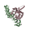
| Title | Crystal structure of Apo form of Short Prokaryotic Argonaute TIR-APAZ (SPARTA) heterodimer | ||||||
 Components Components |
| ||||||
 Keywords Keywords | RNA BINDING PROTEIN / SPARTA / Short Prokaryotic Argonaute / TIR / APAZ | ||||||
| Function / homology |  Function and homology information Function and homology information | ||||||
| Biological species |  Thermoflavifilum thermophilum (bacteria) Thermoflavifilum thermophilum (bacteria) | ||||||
| Method |  X-RAY DIFFRACTION / X-RAY DIFFRACTION /  SYNCHROTRON / SYNCHROTRON /  MOLECULAR REPLACEMENT / Resolution: 2.66 Å MOLECULAR REPLACEMENT / Resolution: 2.66 Å | ||||||
 Authors Authors | Kottur, J. / Aggarwal, A.K. | ||||||
| Funding support | 1items
| ||||||
 Citation Citation |  Journal: Nat Commun / Year: 2024 Journal: Nat Commun / Year: 2024Title: Nucleic acid mediated activation of a short prokaryotic Argonaute immune system. Authors: Jithesh Kottur / Radhika Malik / Aneel K Aggarwal /   Abstract: A short prokaryotic Argonaute (pAgo) TIR-APAZ (SPARTA) defense system, activated by invading DNA to unleash its TIR domain for NAD(P) hydrolysis, was recently identified in bacteria. We report the ...A short prokaryotic Argonaute (pAgo) TIR-APAZ (SPARTA) defense system, activated by invading DNA to unleash its TIR domain for NAD(P) hydrolysis, was recently identified in bacteria. We report the crystal structure of SPARTA heterodimer in the absence of guide-RNA/target-ssDNA (2.66 Å) and a cryo-EM structure of the SPARTA oligomer (tetramer of heterodimers) bound to guide-RNA/target-ssDNA at nominal 3.15-3.35 Å resolution. The crystal structure provides a high-resolution view of SPARTA, revealing the APAZ domain as equivalent to the N, L1, and L2 regions of long pAgos and the MID domain containing a unique insertion (insert57). Cryo-EM structure reveals regions of the PIWI (loop10-9) and APAZ (helix αN) domains that reconfigure for nucleic-acid binding and decrypts regions/residues that reorganize to expose a positively charged pocket for higher-order assembly. The TIR domains amass in a parallel-strands arrangement for catalysis. We visualize SPARTA before and after RNA/ssDNA binding and uncover the basis of its active assembly leading to abortive infection. | ||||||
| History |
|
- Structure visualization
| Structure viewer | Molecule:  Molmil Molmil Jmol/JSmol Jmol/JSmol |
|---|
- Downloads & links
- Download
| PDBx/mmCIF format |  8u7b.cif.gz 8u7b.cif.gz | 396.9 KB | Display |  PDBx/mmCIF format PDBx/mmCIF format |
|---|---|---|---|---|
| PDB format |  pdb8u7b.ent.gz pdb8u7b.ent.gz | 321.3 KB | Display |  PDB format PDB format |
| PDBx/mmJSON format |  8u7b.json.gz 8u7b.json.gz | Tree view |  PDBx/mmJSON format PDBx/mmJSON format | |
| Others |  Other downloads Other downloads |
-Validation report
| Summary document |  8u7b_validation.pdf.gz 8u7b_validation.pdf.gz | 462.3 KB | Display |  wwPDB validaton report wwPDB validaton report |
|---|---|---|---|---|
| Full document |  8u7b_full_validation.pdf.gz 8u7b_full_validation.pdf.gz | 481.4 KB | Display | |
| Data in XML |  8u7b_validation.xml.gz 8u7b_validation.xml.gz | 34.7 KB | Display | |
| Data in CIF |  8u7b_validation.cif.gz 8u7b_validation.cif.gz | 47.6 KB | Display | |
| Arichive directory |  https://data.pdbj.org/pub/pdb/validation_reports/u7/8u7b https://data.pdbj.org/pub/pdb/validation_reports/u7/8u7b ftp://data.pdbj.org/pub/pdb/validation_reports/u7/8u7b ftp://data.pdbj.org/pub/pdb/validation_reports/u7/8u7b | HTTPS FTP |
-Related structure data
| Related structure data |  8u72C C: citing same article ( |
|---|---|
| Similar structure data | Similarity search - Function & homology  F&H Search F&H Search |
- Links
Links
- Assembly
| Deposited unit | 
| ||||||||
|---|---|---|---|---|---|---|---|---|---|
| 1 |
| ||||||||
| Unit cell |
|
- Components
| #1: Protein | Mass: 53141.648 Da / Num. of mol.: 1 Source method: isolated from a genetically manipulated source Source: (gene. exp.)  Thermoflavifilum thermophilum (bacteria) Thermoflavifilum thermophilum (bacteria)Gene: SAMN05660895_1670 / Production host:  | ||||||
|---|---|---|---|---|---|---|---|
| #2: Protein | Mass: 58304.848 Da / Num. of mol.: 1 Source method: isolated from a genetically manipulated source Source: (gene. exp.)  Thermoflavifilum thermophilum (bacteria) Thermoflavifilum thermophilum (bacteria)Gene: SAMN05660895_1671 / Production host:  | ||||||
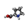
| #3: Chemical | ChemComp-PRO / #4: Chemical | #5: Water | ChemComp-HOH / | Has ligand of interest | N | |
-Experimental details
-Experiment
| Experiment | Method:  X-RAY DIFFRACTION / Number of used crystals: 1 X-RAY DIFFRACTION / Number of used crystals: 1 |
|---|
- Sample preparation
| Crystal | Density Matthews: 4.61 Å3/Da / Density % sol: 73.34 % |
|---|---|
| Crystal grow | Temperature: 293.15 K / Method: vapor diffusion, hanging drop / pH: 4.5 Details: 0.1 M sodium acetate, pH 4.5, 0.8 M sodium phosphate monobasic, 1.2 M potassium phosphate dibasic PH range: 4-6 |
-Data collection
| Diffraction | Mean temperature: 100 K / Serial crystal experiment: N |
|---|---|
| Diffraction source | Source:  SYNCHROTRON / Site: SYNCHROTRON / Site:  NSLS-II NSLS-II  / Beamline: 17-ID-2 / Wavelength: 0.979338 Å / Beamline: 17-ID-2 / Wavelength: 0.979338 Å |
| Detector | Type: DECTRIS EIGER X 16M / Detector: PIXEL / Date: Jul 29, 2022 |
| Radiation | Protocol: SINGLE WAVELENGTH / Monochromatic (M) / Laue (L): M / Scattering type: x-ray |
| Radiation wavelength | Wavelength: 0.979338 Å / Relative weight: 1 |
| Reflection | Resolution: 2.66→170.76 Å / Num. obs: 40914 / % possible obs: 96.2 % / Redundancy: 13.3 % / CC1/2: 0.999 / Rmerge(I) obs: 0.148 / Rpim(I) all: 0.042 / Net I/σ(I): 13.7 |
| Reflection shell | Resolution: 2.66→2.99 Å / Rmerge(I) obs: 1.767 / Mean I/σ(I) obs: 1.7 / Num. unique obs: 2047 / CC1/2: 0.572 / Rpim(I) all: 0.554 |
- Processing
| Software |
| ||||||||||||||||||||||||||||||||||||||||||||||||||||||||||||||||||||||||||||||||||||||||||||||||||||||||||||||||
|---|---|---|---|---|---|---|---|---|---|---|---|---|---|---|---|---|---|---|---|---|---|---|---|---|---|---|---|---|---|---|---|---|---|---|---|---|---|---|---|---|---|---|---|---|---|---|---|---|---|---|---|---|---|---|---|---|---|---|---|---|---|---|---|---|---|---|---|---|---|---|---|---|---|---|---|---|---|---|---|---|---|---|---|---|---|---|---|---|---|---|---|---|---|---|---|---|---|---|---|---|---|---|---|---|---|---|---|---|---|---|---|---|---|
| Refinement | Method to determine structure:  MOLECULAR REPLACEMENT / Resolution: 2.66→49.71 Å / SU ML: 0.27 / Cross valid method: FREE R-VALUE / σ(F): 1.35 / Phase error: 29.72 / Stereochemistry target values: ML MOLECULAR REPLACEMENT / Resolution: 2.66→49.71 Å / SU ML: 0.27 / Cross valid method: FREE R-VALUE / σ(F): 1.35 / Phase error: 29.72 / Stereochemistry target values: ML
| ||||||||||||||||||||||||||||||||||||||||||||||||||||||||||||||||||||||||||||||||||||||||||||||||||||||||||||||||
| Solvent computation | Shrinkage radii: 0.9 Å / VDW probe radii: 1.11 Å / Solvent model: FLAT BULK SOLVENT MODEL | ||||||||||||||||||||||||||||||||||||||||||||||||||||||||||||||||||||||||||||||||||||||||||||||||||||||||||||||||
| Refinement step | Cycle: LAST / Resolution: 2.66→49.71 Å
| ||||||||||||||||||||||||||||||||||||||||||||||||||||||||||||||||||||||||||||||||||||||||||||||||||||||||||||||||
| Refine LS restraints |
| ||||||||||||||||||||||||||||||||||||||||||||||||||||||||||||||||||||||||||||||||||||||||||||||||||||||||||||||||
| LS refinement shell |
|
 Movie
Movie Controller


 PDBj
PDBj


