 Movie
Movie Controller

[English] 日本語
 Yorodumi
- PDB-8u5b: Cryo-EM structure of human claudin-4 complex with Clostridium per... -
+ Open data

- Basic information
| Entry | Database: PDB / ID: 8u5b | ||||||
|---|---|---|---|---|---|---|---|
| Title | Cryo-EM structure of human claudin-4 complex with Clostridium perfringens enterotoxin C-terminal domain and sFab COP-1 | ||||||
 Components Components |
| ||||||
 Keywords Keywords | MEMBRANE PROTEIN / Claudin / Fab / Toxin | ||||||
| Function / homology |  Function and homology information Function and homology informationparacellular transport / calcium-independent cell-cell adhesion / Tight junction interactions / apicolateral plasma membrane / bicellular tight junction assembly / regulation of cell morphogenesis / tight junction / positive regulation of wound healing / renal absorption / chloride channel activity ...paracellular transport / calcium-independent cell-cell adhesion / Tight junction interactions / apicolateral plasma membrane / bicellular tight junction assembly / regulation of cell morphogenesis / tight junction / positive regulation of wound healing / renal absorption / chloride channel activity / establishment of skin barrier / lateral plasma membrane / bicellular tight junction / chloride channel complex / response to progesterone / basal plasma membrane / female pregnancy / circadian rhythm / cell-cell junction / transmembrane signaling receptor activity / toxin activity / cell adhesion / apical plasma membrane / positive regulation of cell migration / structural molecule activity / extracellular region / identical protein binding / plasma membrane Similarity search - Function | ||||||
| Biological species |  Homo sapiens (human) Homo sapiens (human)  Clostridium perfringens (bacteria) Clostridium perfringens (bacteria)synthetic construct (others) | ||||||
| Method | ELECTRON MICROSCOPY / single particle reconstruction / cryo EM / Resolution: 5.3 Å | ||||||
 Authors Authors | Vecchio, A.J. | ||||||
| Funding support |  United States, 1items United States, 1items
| ||||||
 Citation Citation | Journal: Commun Biol / Year: 2024 Title: Structural and biophysical insights into targeting of claudin-4 by a synthetic antibody fragment. Authors: Satchal K Erramilli / Pawel K Dominik / Chinemerem P Ogbu / Anthony A Kossiakoff / Alex J Vecchio / Abstract: Claudins are a 27-member family of ~25 kDa membrane proteins that integrate into tight junctions to form molecular barriers at the paracellular spaces between endothelial and epithelial cells. As ...Claudins are a 27-member family of ~25 kDa membrane proteins that integrate into tight junctions to form molecular barriers at the paracellular spaces between endothelial and epithelial cells. As the backbone of tight junction structure and function, claudins are attractive targets for modulating tissue permeability to deliver drugs or treat disease. However, structures of claudins are limited due to their small sizes and physicochemical properties-these traits also make therapy development a challenge. Here we report the development of a synthetic antibody fragment (sFab) that binds human claudin-4 and the determination of a high-resolution structure of it bound to claudin-4/enterotoxin complexes using cryogenic electron microscopy. Structural and biophysical results reveal this sFabs mechanism of select binding to human claudin-4 over other homologous claudins and establish the ability of sFabs to bind hard-to-target claudins to probe tight junction structure and function. The findings provide a framework for tight junction modulation by sFabs for tissue-selective therapies. #1: Journal: bioRxiv / Year: 2023Title: Cryo-EM structures of a synthetic antibody against 22 kDa claudin-4 reveal its complex with Authors: Erramilli, S.K. / Dominik, P.K. / Ogbu, C.P. / Kossiakoff, A.A. / Vecchio, A.J. | ||||||
| History |
|
- Structure visualization
| Structure viewer | Molecule: MolmilJmol/JSmol |
|---|
- Downloads & links
-Download
| PDBx/mmCIF format | 8u5b.cif.gz | 159.5 KB | Display | PDBx/mmCIF format |
|---|---|---|---|---|
| PDB format | pdb8u5b.ent.gz | 120.4 KB | Display | PDB format |
| PDBx/mmJSON format | 8u5b.json.gz | Tree view | PDBx/mmJSON format | |
| Others |  Other downloads Other downloads |
-Validation report
| Arichive directory | https://data.pdbj.org/pub/pdb/validation_reports/u5/8u5bftp://data.pdbj.org/pub/pdb/validation_reports/u5/8u5b | HTTPS FTP |
|---|
-Related structure data
| Related structure data |  41915MC  8u4vC C: citing same article ( M: map data used to model this data |
|---|---|
| Similar structure data |


-Links
 PDBj
PDBj


- Assembly
| Deposited unit | 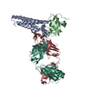
|
|---|---|
| 1 |
|
-Components
| #1: Protein | Mass: 22613.852 Da / Num. of mol.: 1 Source method: isolated from a genetically manipulated source Source: (gene. exp.) Homo sapiens (human) / Gene: CLDN4, CPER, CPETR1, WBSCR8 / Plasmid: pFastBac1 / Cell line (production host): Tn5 / Production host:  Trichoplusia ni (cabbage looper) / References: UniProt: O14493 Trichoplusia ni (cabbage looper) / References: UniProt: O14493 |
|---|---|
| #2: Protein | Mass: 14044.594 Da / Num. of mol.: 1 Source method: isolated from a genetically manipulated source Source: (gene. exp.) Clostridium perfringens (bacteria) / Gene: cpe / Plasmid: pFastBac1 / Cell line (production host): Tn5 / Production host: Trichoplusia ni (cabbage looper) / References: UniProt: P01558 |
| #3: Antibody | Mass: 23258.783 Da / Num. of mol.: 1 Source method: isolated from a genetically manipulated source Source: (gene. exp.) synthetic construct (others) / Production host: |
| #4: Antibody | Mass: 28064.498 Da / Num. of mol.: 1 Source method: isolated from a genetically manipulated source Source: (gene. exp.) synthetic construct (others) / Production host: |
| #5: Chemical | ChemComp-AV0 /   Mass: 1005.188 Da / Num. of mol.: 1 / Source method: obtained synthetically / Formula: C47H88O22 Mass: 1005.188 Da / Num. of mol.: 1 / Source method: obtained synthetically / Formula: C47H88O22 |
| Has ligand of interest | N |
| Has protein modification | Y |
-Experimental details
-Experiment
| Experiment | Method: ELECTRON MICROSCOPY |
|---|---|
| EM experiment | Aggregation state: PARTICLE / 3D reconstruction method: single particle reconstruction |
- Sample preparation
| Component | Name: Human claudin-4 complex with Clostridium perfringens enterotoxin C-terminal domain and sFab COP-1 Type: COMPLEX Details: Assembled complex of 4 proteins (Fab is 2 proteins) expressed from insect cells and E coli Entity ID: #1-#4 / Source: MULTIPLE SOURCES |
|---|---|
| Molecular weight | Value: 0.1 MDa / Experimental value: NO |
| Source (natural) | Organism: Homo sapiens (human) |
| Source (recombinant) | Organism: Trichoplusia ni (cabbage looper) |
| Buffer solution | pH: 7.4 / Details: 20 mM Hepes pH 7.4, 100 mM NaCl, and 0.003% LMNG |
| Specimen | Conc.: 6 mg/ml / Embedding applied: NO / Shadowing applied: NO / Staining applied: NO / Vitrification applied: YES |
| Specimen support | Grid material: COPPER / Grid mesh size: 200 divisions/in. / Grid type: Quantifoil R2/1 |
| Vitrification | Instrument: FEI VITROBOT MARK IV / Cryogen name: ETHANE / Humidity: 100 % / Chamber temperature: 278 K |
- Electron microscopy imaging
| Experimental equipment |  Model: Titan Krios / Image courtesy: FEI Company |
|---|---|
| Microscopy | Model: FEI TITAN KRIOS |
| Electron gun | Electron source:  FIELD EMISSION GUN / Accelerating voltage: 300 kV / Illumination mode: FLOOD BEAM FIELD EMISSION GUN / Accelerating voltage: 300 kV / Illumination mode: FLOOD BEAM |
| Electron lens | Mode: BRIGHT FIELD / Nominal magnification: 92000 X / Nominal defocus max: 2200 nm / Nominal defocus min: 800 nm |
| Specimen holder | Cryogen: NITROGEN / Specimen holder model: FEI TITAN KRIOS AUTOGRID HOLDER |
| Image recording | Electron dose: 60 e/Å2 / Film or detector model: GATAN K3 (6k x 4k) / Num. of grids imaged: 1 / Num. of real images: 1073 |
| EM imaging optics | Energyfilter name: GIF Bioquantum |
- Processing
| EM software |
| ||||||||||||||||||||||||||||||||||||||||
|---|---|---|---|---|---|---|---|---|---|---|---|---|---|---|---|---|---|---|---|---|---|---|---|---|---|---|---|---|---|---|---|---|---|---|---|---|---|---|---|---|---|
| CTF correction | Type: PHASE FLIPPING AND AMPLITUDE CORRECTION | ||||||||||||||||||||||||||||||||||||||||
| Particle selection | Num. of particles selected: 37586 | ||||||||||||||||||||||||||||||||||||||||
| Symmetry | Point symmetry: C1 (asymmetric) | ||||||||||||||||||||||||||||||||||||||||
| 3D reconstruction | Resolution: 5.3 Å / Resolution method: FSC 0.143 CUT-OFF / Num. of particles: 37296 / Algorithm: FOURIER SPACE / Num. of class averages: 1 / Symmetry type: POINT | ||||||||||||||||||||||||||||||||||||||||
| Atomic model building | Protocol: FLEXIBLE FIT / Space: REAL | ||||||||||||||||||||||||||||||||||||||||
| Atomic model building | PDB-ID: 7kp4 Accession code: 7kp4 / Source name: PDB / Type: experimental model | ||||||||||||||||||||||||||||||||||||||||
| Refine LS restraints |
|

