protein import into peroxisome matrix, receptor recycling / protein import into peroxisome matrix / protein transporter activity / peroxisomal membrane / ATPase complex / protein unfolding / Hydrolases; Acting on acid anhydrides; Acting on acid anhydrides to facilitate cellular and subcellular movement / peroxisome / ATP hydrolysis activity / ATP binding / cytosol Similarity search - Function
: / : / PEX6 N1 domain / PEX6 fourth domain / : / AAA ATPase, AAA+ lid domain / AAA+ lid domain / ATPase, AAA-type, conserved site / AAA-protein family signature. / ATPase family associated with various cellular activities (AAA) ...: / : / PEX6 N1 domain / PEX6 fourth domain / : / AAA ATPase, AAA+ lid domain / AAA+ lid domain / ATPase, AAA-type, conserved site / AAA-protein family signature. / ATPase family associated with various cellular activities (AAA) / ATPase, AAA-type, core / ATPases associated with a variety of cellular activities / AAA+ ATPase domain / P-loop containing nucleoside triphosphate hydrolase Similarity search - Domain/homology
National Institutes of Health/National Institute of General Medical Sciences (NIH/NIGMS)
R00GM121880
United States
National Institutes of Health/National Institute of General Medical Sciences (NIH/NIGMS)
R01GM094497
United States
Citation
Journal: J Biol Chem / Year: 2024 Title: The N1 domain of the peroxisomal AAA-ATPase Pex6 is required for Pex15 binding and proper assembly with Pex1. Authors: Bashir A Ali / Ryan M Judy / Saikat Chowdhury / Nicole K Jacobsen / Dominic T Castanzo / Kaili L Carr / Chris D Richardson / Gabriel C Lander / Andreas Martin / Brooke M Gardner / Abstract: The heterohexameric ATPases associated with diverse cellular activities (AAA)-ATPase Pex1/Pex6 is essential for the formation and maintenance of peroxisomes. Pex1/Pex6, similar to other AAA-ATPases, ...The heterohexameric ATPases associated with diverse cellular activities (AAA)-ATPase Pex1/Pex6 is essential for the formation and maintenance of peroxisomes. Pex1/Pex6, similar to other AAA-ATPases, uses the energy from ATP hydrolysis to mechanically thread substrate proteins through its central pore, thereby unfolding them. In related AAA-ATPase motors, substrates are recruited through binding to the motor's N-terminal domains or N terminally bound cofactors. Here, we use structural and biochemical techniques to characterize the function of the N1 domain in Pex6 from budding yeast, Saccharomyces cerevisiae. We found that although Pex1/ΔN1-Pex6 is an active ATPase in vitro, it does not support Pex1/Pex6 function at the peroxisome in vivo. An X-ray crystal structure of the isolated Pex6 N1 domain shows that the Pex6 N1 domain shares the same fold as the N-terminal domains of PEX1, CDC48, and NSF, despite poor sequence conservation. Integrating this structure with a cryo-EM reconstruction of Pex1/Pex6, AlphaFold2 predictions, and biochemical assays shows that Pex6 N1 mediates binding to both the peroxisomal membrane tether Pex15 and an extended loop from the D2 ATPase domain of Pex1 that influences Pex1/Pex6 heterohexamer stability. Given the direct interactions with both Pex15 and the D2 ATPase domains, the Pex6 N1 domain is poised to coordinate binding of cofactors and substrates with Pex1/Pex6 ATPase activity.
PeroxisomalATPasePEX6 / Peroxin-6 / Peroxisomal assembly protein 8
Mass: 20908.201 Da / Num. of mol.: 1 Source method: isolated from a genetically manipulated source Source: (gene. exp.) Saccharomyces cerevisiae (brewer's yeast) Gene: PEX6, PAS8, YNL329C, N0310 / Production host: Escherichia coli (E. coli) References: UniProt: P33760, Hydrolases; Acting on acid anhydrides; Acting on acid anhydrides to facilitate cellular and subcellular movement
In the structure databanks used in Yorodumi, some data are registered as the other names, "COVID-19 virus" and "2019-nCoV". Here are the details of the virus and the list of structure data.
Jan 31, 2019. EMDB accession codes are about to change! (news from PDBe EMDB page)
EMDB accession codes are about to change! (news from PDBe EMDB page)
The allocation of 4 digits for EMDB accession codes will soon come to an end. Whilst these codes will remain in use, new EMDB accession codes will include an additional digit and will expand incrementally as the available range of codes is exhausted. The current 4-digit format prefixed with “EMD-” (i.e. EMD-XXXX) will advance to a 5-digit format (i.e. EMD-XXXXX), and so on. It is currently estimated that the 4-digit codes will be depleted around Spring 2019, at which point the 5-digit format will come into force.
The EM Navigator/Yorodumi systems omit the EMD- prefix.
Related info.:Q: What is EMD? / ID/Accession-code notation in Yorodumi/EM Navigator
Yorodumi is a browser for structure data from EMDB, PDB, SASBDB, etc.
This page is also the successor to EM Navigator detail page, and also detail information page/front-end page for Omokage search.
The word "yorodu" (or yorozu) is an old Japanese word meaning "ten thousand". "mi" (miru) is to see.
Related info.:EMDB / PDB / SASBDB / Comparison of 3 databanks / Yorodumi Search / Aug 31, 2016. New EM Navigator & Yorodumi / Yorodumi Papers / Jmol/JSmol / Function and homology information / Changes in new EM Navigator and Yorodumi
 Movie
Movie Controller


 Open data
 Basic information
 Components
 Keywords
 Function and homology information
 X-RAY DIFFRACTION /
  Authors
 United States, 2items
  Citation
 Structure visualization
 Downloads & links
 Other downloads

 PDBj
PDBj


 Assembly
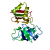

 Mass: 18.015 Da / Num. of mol.: 91 / Source method: isolated from a natural source / Formula: H2O
 Sample preparation
 Processing