 Movie
Movie Controller

[English] 日本語
 Yorodumi
- PDB-8u0m: Crystal structure of isopentenyl phosphate kinase from Thermococc... -
+ Open data

- Basic information
| Entry | Database: PDB / ID: 8u0m | ||||||
|---|---|---|---|---|---|---|---|
| Title | Crystal structure of isopentenyl phosphate kinase from Thermococcus paralvinellae bound to (E)-2-methylbut-2-en-1-yl monophosphate and ATP | ||||||
 Components Components | Isopentenyl phosphate kinase | ||||||
 Keywords Keywords | TRANSFERASE / Alternate Mevalonate pathway IPK Amino acid kinase isoprenoids | ||||||
| Function / homology |  Function and homology information Function and homology informationisopentenyl phosphate kinase / isopentenyl phosphate kinase activity / terpenoid biosynthetic process / kinase activity / ATP binding / cytosol Similarity search - Function | ||||||
| Biological species |   Thermococcus paralvinellae (archaea) Thermococcus paralvinellae (archaea) | ||||||
| Method |  X-RAY DIFFRACTION / SYNCHROTRON / MOLECULAR REPLACEMENT / Resolution: 2.54 Å X-RAY DIFFRACTION / SYNCHROTRON / MOLECULAR REPLACEMENT / Resolution: 2.54 Å | ||||||
 Authors Authors | Singh, S. / Thomas, L.M. / Johnson, B.P. / Brown, S. | ||||||
| Funding support |  United States, 1items United States, 1items
| ||||||
 Citation Citation | Journal: Proteins / Year: 2024 Title: Ternary complexes of isopentenyl phosphate kinase from Thermococcus paralvinellae reveal molecular determinants of non-natural substrate specificity. Authors: Johnson, B.P. / Mandal, P.S. / Brown, S.M. / Thomas, L.M. / Singh, S. | ||||||
| History |
|
- Structure visualization
| Structure viewer | Molecule: MolmilJmol/JSmol |
|---|
- Downloads & links
-Download
| PDBx/mmCIF format | 8u0m.cif.gz | 138.1 KB | Display | PDBx/mmCIF format |
|---|---|---|---|---|
| PDB format | pdb8u0m.ent.gz | 85.9 KB | Display | PDB format |
| PDBx/mmJSON format | 8u0m.json.gz | Tree view | PDBx/mmJSON format | |
| Others |  Other downloads Other downloads |
-Validation report
| Arichive directory | https://data.pdbj.org/pub/pdb/validation_reports/u0/8u0mftp://data.pdbj.org/pub/pdb/validation_reports/u0/8u0m | HTTPS FTP |
|---|
-Related structure data
| Related structure data | 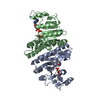 8u0kC 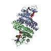 8u0lC 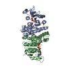 8u0nC 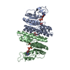 7lntS S: Starting model for refinement C: citing same article ( |
|---|---|
| Similar structure data |
-Links
 PDBj
PDBj- Assembly
| Deposited unit | 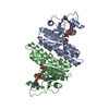
| ||||||||||||
|---|---|---|---|---|---|---|---|---|---|---|---|---|---|
| 1 |
| ||||||||||||
| Unit cell |
| ||||||||||||
| Components on special symmetry positions |
|
-Components
| #1: Protein | Mass: 31962.855 Da / Num. of mol.: 2 Source method: isolated from a genetically manipulated source Source: (gene. exp.) Thermococcus paralvinellae (archaea) / Gene: TES1_0236 / Production host:  #2: Chemical | ChemComp-ADP / |   Mass: 427.201 Da / Num. of mol.: 1 / Source method: obtained synthetically / Formula: C10H15N5O10P2 / Feature type: SUBJECT OF INVESTIGATION / Comment: ADP, energy-carrying molecule*YM Mass: 427.201 Da / Num. of mol.: 1 / Source method: obtained synthetically / Formula: C10H15N5O10P2 / Feature type: SUBJECT OF INVESTIGATION / Comment: ADP, energy-carrying molecule*YM#3: Chemical | Mass: 166.112 Da / Num. of mol.: 2 / Source method: obtained synthetically / Formula: C5H11O4P / Feature type: SUBJECT OF INVESTIGATION #4: Chemical | ChemComp-ATP / |   Mass: 507.181 Da / Num. of mol.: 1 / Source method: obtained synthetically / Formula: C10H16N5O13P3 / Feature type: SUBJECT OF INVESTIGATION / Comment: ATP, energy-carrying molecule*YM Mass: 507.181 Da / Num. of mol.: 1 / Source method: obtained synthetically / Formula: C10H16N5O13P3 / Feature type: SUBJECT OF INVESTIGATION / Comment: ATP, energy-carrying molecule*YM#5: Water | ChemComp-HOH / |  Mass: 18.015 Da / Num. of mol.: 18 / Source method: isolated from a natural source / Formula: H2O Mass: 18.015 Da / Num. of mol.: 18 / Source method: isolated from a natural source / Formula: H2OHas ligand of interest | Y | |
|---|
-Experimental details
-Experiment
| Experiment | Method: X-RAY DIFFRACTION / Number of used crystals: 1 |
|---|
- Sample preparation
| Crystal | Density Matthews: 2.47 Å3/Da / Density % sol: 50.22 % |
|---|---|
| Crystal grow | Temperature: 294 K / Method: vapor diffusion, hanging drop / pH: 7.8 / Details: 0.133 M magnesium formate, 10.0% PEG3350 |
-Data collection
| Diffraction | Mean temperature: 100 K / Serial crystal experiment: N |
|---|---|
| Diffraction source | Source: SYNCHROTRON / Site: SSRL / Beamline: BL9-2 / Wavelength: 0.98 Å |
| Detector | Type: DECTRIS PILATUS 6M / Detector: PIXEL / Date: Feb 13, 2020 |
| Radiation | Protocol: SINGLE WAVELENGTH / Monochromatic (M) / Laue (L): M / Scattering type: x-ray |
| Radiation wavelength | Wavelength: 0.98 Å / Relative weight: 1 |
| Reflection | Resolution: 2.54→40 Å / Num. obs: 19727 / % possible obs: 91 % / Redundancy: 5.9 % / Biso Wilson estimate: 56.19 Å2 / CC1/2: 1 / Rpim(I) all: 0.043 / Rrim(I) all: 0.109 / Net I/σ(I): 15.8 |
| Reflection shell | Resolution: 2.54→2.58 Å / Num. unique obs: 503 / CC1/2: 0.484 / Rpim(I) all: 0.229 / Rrim(I) all: 0.601 / % possible all: 47.5 |
- Processing
| Software |
| |||||||||||||||||||||||||||||||||||||||||||||||||||||||||||||||||||||||||||||||||||||||||||||||||||||||||
|---|---|---|---|---|---|---|---|---|---|---|---|---|---|---|---|---|---|---|---|---|---|---|---|---|---|---|---|---|---|---|---|---|---|---|---|---|---|---|---|---|---|---|---|---|---|---|---|---|---|---|---|---|---|---|---|---|---|---|---|---|---|---|---|---|---|---|---|---|---|---|---|---|---|---|---|---|---|---|---|---|---|---|---|---|---|---|---|---|---|---|---|---|---|---|---|---|---|---|---|---|---|---|---|---|---|---|
| Refinement | Method to determine structure: MOLECULAR REPLACEMENT Starting model: PDB entry 7LNT Resolution: 2.54→39.83 Å / SU ML: 0.3419 / Cross valid method: FREE R-VALUE / σ(F): 1.37 / Phase error: 30.8588 Stereochemistry target values: GeoStd + Monomer Library + CDL v1.2
| |||||||||||||||||||||||||||||||||||||||||||||||||||||||||||||||||||||||||||||||||||||||||||||||||||||||||
| Solvent computation | Shrinkage radii: 0.9 Å / VDW probe radii: 1.11 Å / Solvent model: FLAT BULK SOLVENT MODEL | |||||||||||||||||||||||||||||||||||||||||||||||||||||||||||||||||||||||||||||||||||||||||||||||||||||||||
| Displacement parameters | Biso mean: 61.86 Å2 | |||||||||||||||||||||||||||||||||||||||||||||||||||||||||||||||||||||||||||||||||||||||||||||||||||||||||
| Refinement step | Cycle: LAST / Resolution: 2.54→39.83 Å
| |||||||||||||||||||||||||||||||||||||||||||||||||||||||||||||||||||||||||||||||||||||||||||||||||||||||||
| Refine LS restraints |
| |||||||||||||||||||||||||||||||||||||||||||||||||||||||||||||||||||||||||||||||||||||||||||||||||||||||||
| LS refinement shell |
|
