 Movie
Movie Controller


+ Open data

- Basic information
| Entry | Database: PDB / ID: 8ty1 | |||||||||||||||||||||||||||||||||||||||||||||
|---|---|---|---|---|---|---|---|---|---|---|---|---|---|---|---|---|---|---|---|---|---|---|---|---|---|---|---|---|---|---|---|---|---|---|---|---|---|---|---|---|---|---|---|---|---|---|
| Title | Cryo-EM structure of coagulation factor VIII bound to NB2E9 | |||||||||||||||||||||||||||||||||||||||||||||
 Components Components |
| |||||||||||||||||||||||||||||||||||||||||||||
 Keywords Keywords | BLOOD CLOTTING/INHIBITOR / Coagulation / Inhibitor antibody / factor VIII / BLOOD CLOTTING-INHIBITOR complex | |||||||||||||||||||||||||||||||||||||||||||||
| Function / homology |  Function and homology information Function and homology informationDefective F8 accelerates dissociation of the A2 domain / Defective F8 binding to the cell membrane / Defective F8 secretion / Defective F8 sulfation at Y1699 / Gamma carboxylation, hypusinylation, hydroxylation, and arylsulfatase activation / Defective F8 binding to von Willebrand factor / blood coagulation, intrinsic pathway / platelet alpha granule / Cargo concentration in the ER / COPII-coated ER to Golgi transport vesicle ...Defective F8 accelerates dissociation of the A2 domain / Defective F8 binding to the cell membrane / Defective F8 secretion / Defective F8 sulfation at Y1699 / Gamma carboxylation, hypusinylation, hydroxylation, and arylsulfatase activation / Defective F8 binding to von Willebrand factor / blood coagulation, intrinsic pathway / platelet alpha granule / Cargo concentration in the ER / COPII-coated ER to Golgi transport vesicle / Defective factor IX causes thrombophilia / Defective cofactor function of FVIIIa variant / Defective F9 variant does not activate FX / COPII-mediated vesicle transport / Defective F8 cleavage by thrombin / : / : / endoplasmic reticulum-Golgi intermediate compartment membrane / platelet alpha granule lumen / acute-phase response / Golgi lumen / blood coagulation / Platelet degranulation / oxidoreductase activity / endoplasmic reticulum lumen / copper ion binding / : / extracellular region / plasma membrane Similarity search - Function | |||||||||||||||||||||||||||||||||||||||||||||
| Biological species |   Homo sapiens (human) Homo sapiens (human) | |||||||||||||||||||||||||||||||||||||||||||||
| Method | ELECTRON MICROSCOPY / single particle reconstruction / cryo EM / Resolution: 3.46 Å | |||||||||||||||||||||||||||||||||||||||||||||
 Authors Authors | Childers, K.C. / Spiegel, P.C. | |||||||||||||||||||||||||||||||||||||||||||||
| Funding support |  United States, 2items United States, 2items
| |||||||||||||||||||||||||||||||||||||||||||||
 Citation Citation | Journal: J Thromb Haemost / Year: 2024 Title: Structural basis for inhibition of coagulation factor VIII reveals a shared antigenic hotspot on the C1 domain. Authors: Kenneth C Childers / Ben Cowper / Jordan D Vaughan / Juliet R McGill / Omar Davulcu / Pete Lollar / Christopher B Doering / Carmen H Coxon / Paul C Spiegel /  Abstract: BACKGROUND: Hemophilia A arises from dysfunctional or deficient coagulation factor (F)VIII and leads to inefficient fibrin clot formation and uncontrolled bleeding events. The development of antibody ...BACKGROUND: Hemophilia A arises from dysfunctional or deficient coagulation factor (F)VIII and leads to inefficient fibrin clot formation and uncontrolled bleeding events. The development of antibody inhibitors is a clinical complication in hemophilia A patients receiving FVIII replacement therapy. LE2E9 is an anti-C1 domain inhibitor previously isolated from a mild/moderate hemophilia A patient and disrupts FVIII interactions with von Willebrand factor and FIXa, though the intermolecular contacts that underpin LE2E9-mediated FVIII neutralization are undefined. OBJECTIVES: To determine the structure of the complex between FVIII and LE2E9 and characterize its mechanism of inhibition. METHODS: FVIII was bound to the antigen binding fragment (Fab) of NB2E9, a recombinant construct of LE2E9, and its structure was determined by cryogenic electron microscopy. RESULTS: This report communicates the 3.46 Å structure of FVIII bound to NB2E9, with its epitope comprising FVIII residues S2040 to Y2043, K2065 to W2070, and R2150 to H2155. Structural analysis ...RESULTS: This report communicates the 3.46 Å structure of FVIII bound to NB2E9, with its epitope comprising FVIII residues S2040 to Y2043, K2065 to W2070, and R2150 to H2155. Structural analysis reveals that the LE2E9 epitope overlaps with portions of the epitope for 2A9, a murine-derived inhibitor, suggesting that these residues represent a shared antigenic region on the C1 domain between FVIII mice and hemophilia A patients. Furthermore, the FVIII:NB2E9 structure elucidates the orientation of the LE2E9 glycan, illustrating how the glycan sterically blocks interactions between the FVIII C1 domain and the von Willebrand factor D' domain. A putative model of the FVIIIa:FIXa complex suggests potential clashing between the NB2E9 glycan and FIXa light chain. CONCLUSION: These results describe an antigenic "hotspot" on the FVIII C1 domain and provide a structural basis for engineering FVIII replacement therapeutics with reduced antigenicity. | |||||||||||||||||||||||||||||||||||||||||||||
| History |
|
- Structure visualization
| Structure viewer | Molecule: MolmilJmol/JSmol |
|---|
- Downloads & links
-Download
| PDBx/mmCIF format | 8ty1.cif.gz | 356.1 KB | Display | PDBx/mmCIF format |
|---|---|---|---|---|
| PDB format | pdb8ty1.ent.gz | 277.2 KB | Display | PDB format |
| PDBx/mmJSON format | 8ty1.json.gz | Tree view | PDBx/mmJSON format | |
| Others |  Other downloads Other downloads |
-Validation report
| Arichive directory | https://data.pdbj.org/pub/pdb/validation_reports/ty/8ty1ftp://data.pdbj.org/pub/pdb/validation_reports/ty/8ty1 | HTTPS FTP |
|---|
-Related structure data
| Related structure data |  41710MC M: map data used to model this data C: citing same article ( |
|---|---|
| Similar structure data |
-Links
 PDBj
PDBj


- Assembly
| Deposited unit | 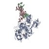
|
|---|---|
| 1 |
|
-Components
| #1: Protein | Mass: 168287.047 Da / Num. of mol.: 1 Source method: isolated from a genetically manipulated source Source: (gene. exp.) Homo sapiens (human)Gene: F8, F8, F8C, F8, CF8 / Production host:  Cricetulus griseus (Chinese hamster) / References: UniProt: P12263, UniProt: P00451 Cricetulus griseus (Chinese hamster) / References: UniProt: P12263, UniProt: P00451 | ||||
|---|---|---|---|---|---|
| #2: Antibody | Mass: 23513.164 Da / Num. of mol.: 1 Source method: isolated from a genetically manipulated source Source: (gene. exp.) Homo sapiens (human) / Production host: Cricetulus griseus (Chinese hamster) | ||||
| #3: Antibody | Mass: 25373.285 Da / Num. of mol.: 1 Source method: isolated from a genetically manipulated source Source: (gene. exp.) Homo sapiens (human) / Production host: Cricetulus griseus (Chinese hamster) | ||||
| #4: Polysaccharide | Source method: isolated from a genetically manipulated source Has ligand of interest | N | Has protein modification | Y | |
-Experimental details
-Experiment
| Experiment | Method: ELECTRON MICROSCOPY |
|---|---|
| EM experiment | Aggregation state: PARTICLE / 3D reconstruction method: single particle reconstruction |
- Sample preparation
| Component | Name: Coagulation factor VIII bound to NB2E9 / Type: COMPLEX / Entity ID: #1-#3 / Source: MULTIPLE SOURCES | |||||||||||||||
|---|---|---|---|---|---|---|---|---|---|---|---|---|---|---|---|---|
| Molecular weight | Experimental value: NO | |||||||||||||||
| Source (natural) | Organism: Homo sapiens (human) | |||||||||||||||
| Source (recombinant) | Organism: Cricetulus griseus (Chinese hamster) | |||||||||||||||
| Buffer solution | pH: 7.4 / Details: 20 mM HEPES (pH 7.4) and 150 mM NaCl | |||||||||||||||
| Buffer component |
| |||||||||||||||
| Specimen | Conc.: 0.1 mg/ml / Embedding applied: NO / Shadowing applied: NO / Staining applied: NO / Vitrification applied: YES | |||||||||||||||
| Specimen support | Grid mesh size: 400 divisions/in. | |||||||||||||||
| Vitrification | Instrument: FEI VITROBOT MARK IV / Cryogen name: ETHANE / Humidity: 100 % / Chamber temperature: 277 K |
- Electron microscopy imaging
| Experimental equipment |  Model: Titan Krios / Image courtesy: FEI Company |
|---|---|
| Microscopy | Model: TFS KRIOS |
| Electron gun | Electron source:  FIELD EMISSION GUN / Accelerating voltage: 300 kV / Illumination mode: FLOOD BEAM FIELD EMISSION GUN / Accelerating voltage: 300 kV / Illumination mode: FLOOD BEAM |
| Electron lens | Mode: BRIGHT FIELD / Nominal defocus max: 2700 nm / Nominal defocus min: 1000 nm |
| Image recording | Electron dose: 50 e/Å2 / Film or detector model: FEI FALCON III (4k x 4k) |
- Processing
| EM software |
| ||||||||||||||||||||||||
|---|---|---|---|---|---|---|---|---|---|---|---|---|---|---|---|---|---|---|---|---|---|---|---|---|---|
| CTF correction | Type: PHASE FLIPPING AND AMPLITUDE CORRECTION | ||||||||||||||||||||||||
| 3D reconstruction | Resolution: 3.46 Å / Resolution method: FSC 0.143 CUT-OFF / Num. of particles: 94935 / Symmetry type: POINT | ||||||||||||||||||||||||
| Refine LS restraints |
|
