 Movie
Movie Controller


+ Open data

- Basic information
| Entry | Database: PDB / ID: 8txr | |||||||||||||||||||||
|---|---|---|---|---|---|---|---|---|---|---|---|---|---|---|---|---|---|---|---|---|---|---|
| Title | E. coli ExoVII(H238A) | |||||||||||||||||||||
 Components Components |
| |||||||||||||||||||||
 Keywords Keywords | DNA BINDING PROTEIN / Exonuclease / Endonuclease / DNA repair | |||||||||||||||||||||
| Function / homology |  Function and homology information Function and homology informationexodeoxyribonuclease VII / exodeoxyribonuclease VII activity / exodeoxyribonuclease VII complex / DNA catabolic process / mismatch repair / DNA binding / cytosol / cytoplasm Similarity search - Function | |||||||||||||||||||||
| Biological species |  | |||||||||||||||||||||
| Method | ELECTRON MICROSCOPY / single particle reconstruction / cryo EM / Resolution: 3.8 Å | |||||||||||||||||||||
 Authors Authors | Liu, C. / Berger, J.M. | |||||||||||||||||||||
| Funding support |  United States, 1items United States, 1items
| |||||||||||||||||||||
 Citation Citation | Journal: Proc Natl Acad Sci U S A / Year: 2024 Title: Structure of exonuclease VII. Authors: Chuan Liu / Glenn Hauk / Qianyun Yan / James M Berger / Abstract: Exonuclease VII (ExoVII) is a ubiquitous bacterial nuclease. Encoded by the and genes, ExoVII participates in multiple nucleic acid-dependent pathways including the processing of multicopy single- ...Exonuclease VII (ExoVII) is a ubiquitous bacterial nuclease. Encoded by the and genes, ExoVII participates in multiple nucleic acid-dependent pathways including the processing of multicopy single-stranded DNA and the repair of covalent DNA-protein crosslinks (DPCs). Although many biochemical properties of ExoVII have been defined, little is known about its structure/function relationships. Here, we use cryoelectron microscopy (cryoEM) to determine that ExoVII comprises a highly elongated XseA·XseB holo-complex. Each XseA subunit dimerizes through a central extended α-helical segment decorated by six XseB subunits and a C-terminal, domain-swapped β-barrel element; two XseA·XseB subcomplexes further associate using N-terminal OB (oligonucleotide/oligosaccharide-binding) folds and catalytic domains to form a spindle-shaped, catenated octaicosamer. The catalytic domains of XseA, which adopt a nuclease fold related to 3-dehydroquinate dehydratases, are sequestered in the center of the complex and accessible only through large pores formed between XseA tetramers. The architectural organization of ExoVII, combined with biochemical studies, indicate that substrate selectivity is controlled by steric access to its nuclease elements and that tetramer dissociation results from substrate DNA binding. Despite a lack of sequence and fold homology, the physical organization of ExoVII is reminiscent of Mre11·Rad50/SbcCD ATP (adenosine triphosphate)-dependent nucleases used in the repair of double-stranded DNA breaks, including those formed by DPCs through aberrant topoisomerase activity, suggesting that there may have been convergent evolutionary pressure to contend with such damage events. | |||||||||||||||||||||
| History |
|
- Structure visualization
| Structure viewer | Molecule: MolmilJmol/JSmol |
|---|
- Downloads & links
-Download
| PDBx/mmCIF format | 8txr.cif.gz | 416.6 KB | Display | PDBx/mmCIF format |
|---|---|---|---|---|
| PDB format | pdb8txr.ent.gz | 342.7 KB | Display | PDB format |
| PDBx/mmJSON format | 8txr.json.gz | Tree view | PDBx/mmJSON format | |
| Others |  Other downloads Other downloads |
-Validation report
| Arichive directory | https://data.pdbj.org/pub/pdb/validation_reports/tx/8txrftp://data.pdbj.org/pub/pdb/validation_reports/tx/8txr | HTTPS FTP |
|---|
-Related structure data
| Related structure data | 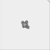 41704MC C: citing same article ( M: map data used to model this data |
|---|---|
| Similar structure data |


-Links
 PDBj
PDBj- Assembly
| Deposited unit | 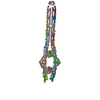
|
|---|---|
| 1 |
|
-Components
| #1: Protein | Mass: 51840.141 Da / Num. of mol.: 4 / Mutation: H238A Source method: isolated from a genetically manipulated source Source: (gene. exp.) Production host: References: UniProt: P04994 #2: Protein | Mass: 8959.877 Da / Num. of mol.: 16 Source method: isolated from a genetically manipulated source Source: (gene. exp.) Production host: References: UniProt: P0A8G9 Has protein modification | N | |
|---|
-Experimental details
-Experiment
| Experiment | Method: ELECTRON MICROSCOPY |
|---|---|
| EM experiment | Aggregation state: PARTICLE / 3D reconstruction method: single particle reconstruction |
- Sample preparation
| Component | Name: XseA4-XseB24 complex of ExoVII / Type: COMPLEX / Entity ID: all / Source: RECOMBINANT |
|---|---|
| Molecular weight | Value: 0.42 MDa / Experimental value: YES |
| Source (natural) | Organism: |
| Source (recombinant) | Organism: |
| Buffer solution | pH: 7.5 |
| Specimen | Embedding applied: NO / Shadowing applied: NO / Staining applied: NO / Vitrification applied: YES |
| Vitrification | Cryogen name: ETHANE |
- Electron microscopy imaging
| Experimental equipment |  Model: Titan Krios / Image courtesy: FEI Company |
|---|---|
| Microscopy | Model: FEI TITAN KRIOS |
| Electron gun | Electron source:  FIELD EMISSION GUN / Accelerating voltage: 300 kV / Illumination mode: FLOOD BEAM FIELD EMISSION GUN / Accelerating voltage: 300 kV / Illumination mode: FLOOD BEAM |
| Electron lens | Mode: BRIGHT FIELD / Nominal defocus max: 1750 nm / Nominal defocus min: 750 nm |
| Image recording | Electron dose: 50.3 e/Å2 / Film or detector model: GATAN K3 BIOQUANTUM (6k x 4k) |
- Processing
| EM software | Name: PHENIX / Category: model refinement | ||||||||||||||||||||||||
|---|---|---|---|---|---|---|---|---|---|---|---|---|---|---|---|---|---|---|---|---|---|---|---|---|---|
| CTF correction | Type: PHASE FLIPPING AND AMPLITUDE CORRECTION | ||||||||||||||||||||||||
| 3D reconstruction | Resolution: 3.8 Å / Resolution method: FSC 0.143 CUT-OFF / Num. of particles: 497337 / Symmetry type: POINT | ||||||||||||||||||||||||
| Refine LS restraints |
|
