National Institutes of Health/National Cancer Institute (NIH/NCI)
United States
Citation
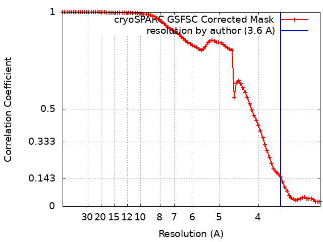
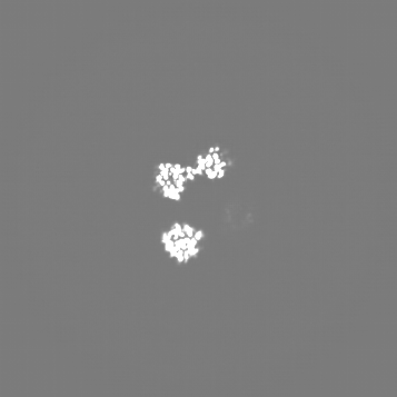
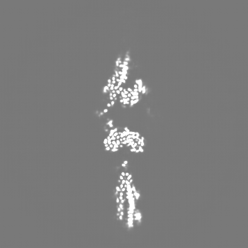
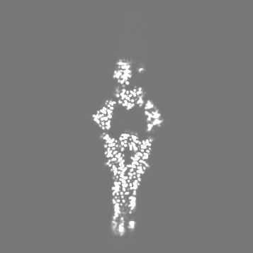
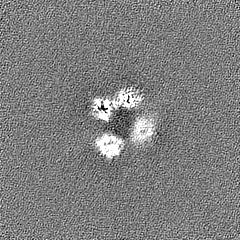
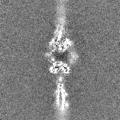
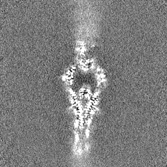
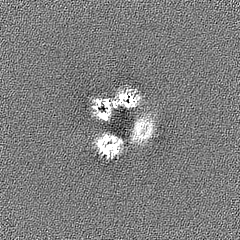
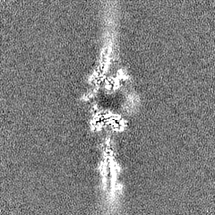
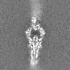
Journal: Proc Natl Acad Sci U S A / Year: 2024 Title: Structure of exonuclease VII. Authors: Chuan Liu / Glenn Hauk / Qianyun Yan / James M Berger / Abstract: Exonuclease VII (ExoVII) is a ubiquitous bacterial nuclease. Encoded by the and genes, ExoVII participates in multiple nucleic acid-dependent pathways including the processing of multicopy single- ...Exonuclease VII (ExoVII) is a ubiquitous bacterial nuclease. Encoded by the and genes, ExoVII participates in multiple nucleic acid-dependent pathways including the processing of multicopy single-stranded DNA and the repair of covalent DNA-protein crosslinks (DPCs). Although many biochemical properties of ExoVII have been defined, little is known about its structure/function relationships. Here, we use cryoelectron microscopy (cryoEM) to determine that ExoVII comprises a highly elongated XseA·XseB holo-complex. Each XseA subunit dimerizes through a central extended α-helical segment decorated by six XseB subunits and a C-terminal, domain-swapped β-barrel element; two XseA·XseB subcomplexes further associate using N-terminal OB (oligonucleotide/oligosaccharide-binding) folds and catalytic domains to form a spindle-shaped, catenated octaicosamer. The catalytic domains of XseA, which adopt a nuclease fold related to 3-dehydroquinate dehydratases, are sequestered in the center of the complex and accessible only through large pores formed between XseA tetramers. The architectural organization of ExoVII, combined with biochemical studies, indicate that substrate selectivity is controlled by steric access to its nuclease elements and that tetramer dissociation results from substrate DNA binding. Despite a lack of sequence and fold homology, the physical organization of ExoVII is reminiscent of Mre11·Rad50/SbcCD ATP (adenosine triphosphate)-dependent nucleases used in the repair of double-stranded DNA breaks, including those formed by DPCs through aberrant topoisomerase activity, suggesting that there may have been convergent evolutionary pressure to contend with such damage events.
In the structure databanks used in Yorodumi, some data are registered as the other names, "COVID-19 virus" and "2019-nCoV". Here are the details of the virus and the list of structure data.
Jan 31, 2019. EMDB accession codes are about to change! (news from PDBe EMDB page)
EMDB accession codes are about to change! (news from PDBe EMDB page)
The allocation of 4 digits for EMDB accession codes will soon come to an end. Whilst these codes will remain in use, new EMDB accession codes will include an additional digit and will expand incrementally as the available range of codes is exhausted. The current 4-digit format prefixed with “EMD-” (i.e. EMD-XXXX) will advance to a 5-digit format (i.e. EMD-XXXXX), and so on. It is currently estimated that the 4-digit codes will be depleted around Spring 2019, at which point the 5-digit format will come into force.
The EM Navigator/Yorodumi systems omit the EMD- prefix.
Related info.:Q: What is EMD? / ID/Accession-code notation in Yorodumi/EM Navigator
Yorodumi is a browser for structure data from EMDB, PDB, SASBDB, etc.
This page is also the successor to EM Navigator detail page, and also detail information page/front-end page for Omokage search.
The word "yorodu" (or yorozu) is an old Japanese word meaning "ten thousand". "mi" (miru) is to see.
Related info.:EMDB / PDB / SASBDB / Comparison of 3 databanks / Yorodumi Search / Aug 31, 2016. New EM Navigator & Yorodumi / Yorodumi Papers / Jmol/JSmol / Function and homology information / Changes in new EM Navigator and Yorodumi
 Movie
Movie Controller


 Open data
 Basic information
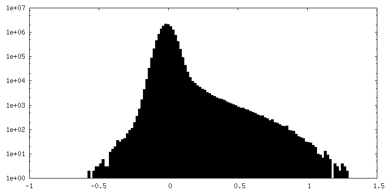
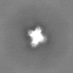
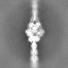
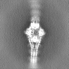
 Map data
 Sample
 Keywords
 Authors
 United States, 1 items
  Citation
 Structure visualization
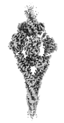
 Downloads & links
 EMDB map data format
 emd_41700.png
 http://ftp.pdbj.org/pub/emdb/structures/EMD-41700

 Z (Sec.)
 Y (Row.)
 X (Col.)


 Sample components
 Processing
 Electron microscopy
 FIELD EMISSION GUN