 Movie
Movie Controller

[English] 日本語
 Yorodumi
- PDB-8tw3: hMPV fusion protein complexed with single domain antibodies sdHMP... -
+ Open data

- Basic information
| Entry | Database: PDB / ID: 8tw3 | |||||||||
|---|---|---|---|---|---|---|---|---|---|---|
| Title | hMPV fusion protein complexed with single domain antibodies sdHMPV16 and sdHMPV12 | |||||||||
 Components Components |
| |||||||||
 Keywords Keywords | VIRAL PROTEIN/IMMUNE SYSTEM / metapneumovirus / fusion protein / VIRAL PROTEIN / VIRAL PROTEIN-Immune System complex | |||||||||
| Function / homology | Precursor fusion glycoprotein F0, Paramyxoviridae / Fusion glycoprotein F0 / fusion of virus membrane with host plasma membrane / viral envelope / symbiont entry into host cell / host cell plasma membrane / virion membrane / Fusion glycoprotein F0 Function and homology information Function and homology information | |||||||||
| Biological species |   Human metapneumovirus A Human metapneumovirus A | |||||||||
| Method |  X-RAY DIFFRACTION / SYNCHROTRON / MOLECULAR REPLACEMENT / Resolution: 2.9 Å X-RAY DIFFRACTION / SYNCHROTRON / MOLECULAR REPLACEMENT / Resolution: 2.9 Å | |||||||||
 Authors Authors | Rush, S.A. / McLellan, J.S. | |||||||||
| Funding support |  United States, 2items United States, 2items
| |||||||||
 Citation Citation | Journal: Mbio / Year: 2024 Title: A neutralizing single-domain antibody that targets the trimer interface of the human metapneumovirus fusion protein. Authors: Ballegeer, M. / van Scherpenzeel, R.C. / Delgado, T. / Iglesias-Caballero, M. / Garcia Barreno, B. / Pandey, S. / Rush, S.A. / Kolkman, J.A. / Mas, V. / McLellan, J.S. / Saelens, X. | |||||||||
| History |
|
- Structure visualization
| Structure viewer | Molecule: MolmilJmol/JSmol |
|---|
- Downloads & links
-Download
| PDBx/mmCIF format | 8tw3.cif.gz | 459.1 KB | Display | PDBx/mmCIF format |
|---|---|---|---|---|
| PDB format | pdb8tw3.ent.gz | 374.1 KB | Display | PDB format |
| PDBx/mmJSON format | 8tw3.json.gz | Tree view | PDBx/mmJSON format | |
| Others |  Other downloads Other downloads |
-Validation report
| Arichive directory | https://data.pdbj.org/pub/pdb/validation_reports/tw/8tw3ftp://data.pdbj.org/pub/pdb/validation_reports/tw/8tw3 | HTTPS FTP |
|---|
-Related structure data
| Similar structure data |
|---|
-Links
 PDBj
PDBj


- Assembly
| Deposited unit | 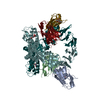
| ||||||||
|---|---|---|---|---|---|---|---|---|---|
| 1 | 
| ||||||||
| 2 | 
| ||||||||
| Unit cell |
|
-Components
| #1: Antibody | Mass: 20633.838 Da / Num. of mol.: 2 Source method: isolated from a genetically manipulated source Source: (gene. exp.)  Homo sapiens (human) Homo sapiens (human)#2: Antibody | Mass: 20863.871 Da / Num. of mol.: 2 Source method: isolated from a genetically manipulated source Source: (gene. exp.) Homo sapiens (human)#3: Protein | Mass: 58805.473 Da / Num. of mol.: 2 Source method: isolated from a genetically manipulated source Source: (gene. exp.) Human metapneumovirus A / Production host: Homo sapiens (human) / References: UniProt: H6X1Z0#4: Sugar | ChemComp-NAG /   Type: D-saccharide, beta linking / Mass: 221.208 Da / Num. of mol.: 4 / Source method: obtained synthetically / Formula: C8H15NO6 Type: D-saccharide, beta linking / Mass: 221.208 Da / Num. of mol.: 4 / Source method: obtained synthetically / Formula: C8H15NO6Has ligand of interest | N | Has protein modification | Y | |
|---|
-Experimental details
-Experiment
| Experiment | Method: X-RAY DIFFRACTION / Number of used crystals: 1 |
|---|
- Sample preparation
| Crystal grow | Temperature: 293 K / Method: vapor diffusion / pH: 7 / Details: 0.1M HEPES and 18% PEG 12,000 |
|---|
-Data collection
| Diffraction | Mean temperature: 100 K / Serial crystal experiment: N |
|---|---|
| Diffraction source | Source: SYNCHROTRON / Site: APS / Beamline: 19-ID / Wavelength: 0.9795 Å |
| Detector | Type: DECTRIS PILATUS 6M / Detector: PIXEL / Date: Dec 2, 2019 |
| Radiation | Protocol: SINGLE WAVELENGTH / Monochromatic (M) / Laue (L): M / Scattering type: x-ray |
| Radiation wavelength | Wavelength: 0.9795 Å / Relative weight: 1 |
| Reflection | Resolution: 2.9→44.97 Å / Num. obs: 32688 / % possible obs: 98.6 % / Redundancy: 5.6 % / CC1/2: 0.962 / Rmerge(I) obs: 0.214 / Rpim(I) all: 0.099 / Rrim(I) all: 0.237 / Χ2: 1.04 / Net I/σ(I): 7 / Num. measured all: 181517 |
| Reflection shell | Resolution: 2.9→3.06 Å / % possible obs: 99.3 % / Redundancy: 5.7 % / Rmerge(I) obs: 0.862 / Num. measured all: 27228 / Num. unique obs: 4780 / CC1/2: 0.638 / Rpim(I) all: 0.393 / Rrim(I) all: 0.951 / Χ2: 0.95 / Net I/σ(I) obs: 2.4 |
- Processing
| Software |
| ||||||||||||||||||||||||||||||||||||||||||||||||||||||||||||||||||||||||||||||||||||
|---|---|---|---|---|---|---|---|---|---|---|---|---|---|---|---|---|---|---|---|---|---|---|---|---|---|---|---|---|---|---|---|---|---|---|---|---|---|---|---|---|---|---|---|---|---|---|---|---|---|---|---|---|---|---|---|---|---|---|---|---|---|---|---|---|---|---|---|---|---|---|---|---|---|---|---|---|---|---|---|---|---|---|---|---|---|
| Refinement | Method to determine structure: MOLECULAR REPLACEMENT / Resolution: 2.9→44.97 Å / SU ML: 0.29 / Cross valid method: FREE R-VALUE / σ(F): 1.33 / Phase error: 22.31 / Stereochemistry target values: ML
| ||||||||||||||||||||||||||||||||||||||||||||||||||||||||||||||||||||||||||||||||||||
| Solvent computation | Shrinkage radii: 0.9 Å / VDW probe radii: 1.1 Å / Solvent model: FLAT BULK SOLVENT MODEL | ||||||||||||||||||||||||||||||||||||||||||||||||||||||||||||||||||||||||||||||||||||
| Refinement step | Cycle: LAST / Resolution: 2.9→44.97 Å
| ||||||||||||||||||||||||||||||||||||||||||||||||||||||||||||||||||||||||||||||||||||
| Refine LS restraints |
| ||||||||||||||||||||||||||||||||||||||||||||||||||||||||||||||||||||||||||||||||||||
| LS refinement shell |
|
