 Movie
Movie Controller


+ Open data

- Basic information
| Entry | Database: PDB / ID: 8tqe | ||||||
|---|---|---|---|---|---|---|---|
| Title | XptA2 wild type | ||||||
 Components Components | XptA2 | ||||||
 Keywords Keywords | TOXIN / TcA / Insecticide / Translocase | ||||||
| Function / homology |  Function and homology information Function and homology informationTcA receptor binding domain / TcA receptor binding domain / ABC toxin, N-terminal domain / ABC toxin N-terminal region / Insecticidal toxin complex/plasmid virulence protein / Tc toxin complex TcA, C-terminal TcB-binding domain / Neuraminidase-like domain / Salmonella virulence plasmid 28.1kDa A protein / Tc toxin complex TcA C-terminal TcB-binding domain / Neuraminidase-like domain Similarity search - Domain/homology | ||||||
| Biological species |  Xenorhabdus nematophila (bacteria) Xenorhabdus nematophila (bacteria) | ||||||
| Method | ELECTRON MICROSCOPY / single particle reconstruction / cryo EM / Resolution: 3.1 Å | ||||||
 Authors Authors | Martin, C.L. / Binshtein, E.M. / Aller, S.G. | ||||||
| Funding support |  United States, 1items United States, 1items
| ||||||
 Citation Citation | Journal: Int J Mol Sci / Year: 2023 Title: Structures of the Insecticidal Toxin Complex Subunit XptA2 Highlight Roles for Flexible Domains. Authors: Cole L Martin / David W Chester / Christopher D Radka / Lurong Pan / Zhengrong Yang / Rachel C Hart / Elad M Binshtein / Zhao Wang / Lisa Nagy / Lawrence J DeLucas / Stephen G Aller / Abstract: The Toxin Complex (Tc) superfamily consists of toxin translocases that contribute to the targeting, delivery, and cytotoxicity of certain pathogenic Gram-negative bacteria. Membrane receptor ...The Toxin Complex (Tc) superfamily consists of toxin translocases that contribute to the targeting, delivery, and cytotoxicity of certain pathogenic Gram-negative bacteria. Membrane receptor targeting is driven by the A-subunit (TcA), which comprises IgG-like receptor binding domains (RBDs) at the surface. To better understand XptA2, an insect specific TcA secreted by the symbiont from the intestine of entomopathogenic nematodes, we determined structures by X-ray crystallography and cryo-EM. Contrary to a previous report, XptA2 is pentameric. RBD-B exhibits an indentation from crystal packing that indicates loose association with the shell and a hotspot for possible receptor binding or a trigger for conformational dynamics. A two-fragment XptA2 lacking an intact linker achieved the folded pre-pore state like wild type (wt), revealing no requirement of the linker for protein folding. The linker is disordered in all structures, and we propose it plays a role in dynamics downstream of the initial pre-pore state. | ||||||
| History |
|
- Structure visualization
| Structure viewer | Molecule: MolmilJmol/JSmol |
|---|
- Downloads & links
-Download
| PDBx/mmCIF format | 8tqe.cif.gz | 2.9 MB | Display | PDBx/mmCIF format |
|---|---|---|---|---|
| PDB format | pdb8tqe.ent.gz | Display | PDB format | |
| PDBx/mmJSON format | 8tqe.json.gz | Tree view | PDBx/mmJSON format | |
| Others |  Other downloads Other downloads |
-Validation report
| Arichive directory | https://data.pdbj.org/pub/pdb/validation_reports/tq/8tqeftp://data.pdbj.org/pub/pdb/validation_reports/tq/8tqe | HTTPS FTP |
|---|
-Related structure data
| Related structure data | 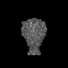 41503MC  8tv0C M: map data used to model this data C: citing same article ( |
|---|---|
| Similar structure data |
-Links
 PDBj
PDBj- Assembly
| Deposited unit | 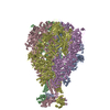
|
|---|---|
| 1 |
|
-Components
| #1: Protein | Mass: 284392.188 Da / Num. of mol.: 5 Source method: isolated from a genetically manipulated source Source: (gene. exp.) Xenorhabdus nematophila (bacteria) / Gene: xptA2 / Production host: |
|---|
-Experimental details
-Experiment
| Experiment | Method: ELECTRON MICROSCOPY |
|---|---|
| EM experiment | Aggregation state: PARTICLE / 3D reconstruction method: single particle reconstruction |
- Sample preparation
| Component | Name: Pentameric assembly of the XptA2 TcA / Type: COMPLEX Details: This is a Protein toxin that is secreted in bacteria. I assume the best category to pick from is Organelle or Cellular Component Entity ID: all / Source: RECOMBINANT |
|---|---|
| Molecular weight | Value: 1.4 MDa / Experimental value: YES |
| Source (natural) | Organism: Xenorhabdus nematophila (bacteria) |
| Source (recombinant) | Organism: |
| Buffer solution | pH: 7.4 |
| Specimen | Conc.: 1.5 mg/ml / Embedding applied: NO / Shadowing applied: NO / Staining applied: NO / Vitrification applied: YES |
| Specimen support | Grid material: COPPER |
| Vitrification | Cryogen name: ETHANE |
- Electron microscopy imaging
| Experimental equipment |  Model: Tecnai Polara / Image courtesy: FEI Company |
|---|---|
| Microscopy | Model: FEI POLARA 300 |
| Electron gun | Electron source:  FIELD EMISSION GUN / Accelerating voltage: 300 kV / Illumination mode: FLOOD BEAM FIELD EMISSION GUN / Accelerating voltage: 300 kV / Illumination mode: FLOOD BEAM |
| Electron lens | Mode: BRIGHT FIELD / Nominal defocus max: 4000 nm / Nominal defocus min: 500 nm / Cs: 2.2 mm / C2 aperture diameter: 50 µm |
| Specimen holder | Cryogen: NITROGEN |
| Image recording | Electron dose: 75 e/Å2 / Detector mode: COUNTING / Film or detector model: GATAN K2 SUMMIT (4k x 4k) |
| Image scans | Movie frames/image: 60 |
- Processing
| EM software |
| ||||||||||||||||||||||||
|---|---|---|---|---|---|---|---|---|---|---|---|---|---|---|---|---|---|---|---|---|---|---|---|---|---|
| CTF correction | Type: PHASE FLIPPING AND AMPLITUDE CORRECTION | ||||||||||||||||||||||||
| Symmetry | Point symmetry: C5 (5 fold cyclic) | ||||||||||||||||||||||||
| 3D reconstruction | Resolution: 3.1 Å / Resolution method: FSC 0.143 CUT-OFF / Num. of particles: 198591 / Symmetry type: POINT | ||||||||||||||||||||||||
| Refinement | Cross valid method: NONE Stereochemistry target values: GeoStd + Monomer Library + CDL v1.2 | ||||||||||||||||||||||||
| Displacement parameters | Biso mean: 47.01 Å2 | ||||||||||||||||||||||||
| Refine LS restraints |
|
