National Institutes of Health/National Institute of General Medical Sciences (NIH/NIGMS)
R35 GM132120
米国
引用
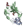
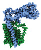
ジャーナル: Nat Commun / 年: 2023 タイトル: Structural basis of peptidoglycan synthesis by E. coli RodA-PBP2 complex. 著者: Rie Nygaard / Chris L B Graham / Meagan Belcher Dufrisne / Jonathan D Colburn / Joseph Pepe / Molly A Hydorn / Silvia Corradi / Chelsea M Brown / Khuram U Ashraf / Owen N Vickery / Nicholas S ...著者: Rie Nygaard / Chris L B Graham / Meagan Belcher Dufrisne / Jonathan D Colburn / Joseph Pepe / Molly A Hydorn / Silvia Corradi / Chelsea M Brown / Khuram U Ashraf / Owen N Vickery / Nicholas S Briggs / John J Deering / Brian Kloss / Bruno Botta / Oliver B Clarke / Linda Columbus / Jonathan Dworkin / Phillip J Stansfeld / David I Roper / Filippo Mancia / 要旨: Peptidoglycan (PG) is an essential structural component of the bacterial cell wall that is synthetized during cell division and elongation. PG forms an extracellular polymer crucial for cellular ...Peptidoglycan (PG) is an essential structural component of the bacterial cell wall that is synthetized during cell division and elongation. PG forms an extracellular polymer crucial for cellular viability, the synthesis of which is the target of many antibiotics. PG assembly requires a glycosyltransferase (GT) to generate a glycan polymer using a Lipid II substrate, which is then crosslinked to the existing PG via a transpeptidase (TP) reaction. A Shape, Elongation, Division and Sporulation (SEDS) GT enzyme and a Class B Penicillin Binding Protein (PBP) form the core of the multi-protein complex required for PG assembly. Here we used single particle cryo-electron microscopy to determine the structure of a cell elongation-specific E. coli RodA-PBP2 complex. We combine this information with biochemical, genetic, spectroscopic, and computational analyses to identify the Lipid II binding sites and propose a mechanism for Lipid II polymerization. Our data suggest a hypothesis for the movement of the glycan strand from the Lipid II polymerization site of RodA towards the TP site of PBP2, functionally linking these two central enzymatic activities required for cell wall peptidoglycan biosynthesis.
 ムービー
ムービー コントローラー


 データを開く
 基本情報
基本情報 要素
要素 キーワード
キーワード 機能・相同性情報
 データ登録者
 米国, 1件
  引用
引用

 構造の表示
構造の表示 ダウンロードとリンク
 その他のダウンロード
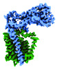

 PDBj
PDBj
 集合体
集合体
 試料調製
試料調製 電子顕微鏡撮影
 FIELD EMISSION GUN / 加速電圧: 300 kV / 照射モード: FLOOD BEAM
 解析
解析