[English] 日本語
Yorodumi
- PDB-8tf5: Crystal structure of orphan G protein-coupled receptor 6, pseudoa... -

+
Open data


ID or keywords:

Loading...

-
Basic information

Entry
Database: PDB / ID: 8tf5
TitleCrystal structure of orphan G protein-coupled receptor 6, pseudoapo form
ComponentsG-protein coupled receptor 6,Soluble cytochrome b562 chimera,Soluble cytochrome b562,G-protein coupled receptor 6
Keywords
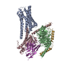
MEMBRANE PROTEIN / Orphan GPCR / GPR6 / pseudoapo form / BRIL / LCP / Parkinson's disease / active-like conformation
Function / homology
Function and homology information


sphingosine-1-phosphate receptor activity / regulation of metabolic process / electron transport chain / G protein-coupled receptor activity / adenylate cyclase-activating G protein-coupled receptor signaling pathway / positive regulation of cytosolic calcium ion concentration / periplasmic space / electron transfer activity / G protein-coupled receptor signaling pathway / iron ion binding ...sphingosine-1-phosphate receptor activity / regulation of metabolic process / electron transport chain / G protein-coupled receptor activity / adenylate cyclase-activating G protein-coupled receptor signaling pathway / positive regulation of cytosolic calcium ion concentration / periplasmic space / electron transfer activity / G protein-coupled receptor signaling pathway / iron ion binding / heme binding / plasma membrane / cytoplasm
Similarity search - Function
G protein-coupled receptor 6 / G protein-coupled receptor 3/6/12 orphan / Cytochrome b562 / Cytochrome b562 / Cytochrome c/b562 / G-protein coupled receptors family 1 signature. / G protein-coupled receptor, rhodopsin-like / GPCR, rhodopsin-like, 7TM / G-protein coupled receptors family 1 profile. / 7 transmembrane receptor (rhodopsin family)
Similarity search - Domain/homology
CHOLESTEROL / OLEIC ACID / (2R)-2,3-dihydroxypropyl (9Z)-octadec-9-enoate / 2,5,8,11,14,17-HEXAOXANONADECAN-19-OL / Soluble cytochrome b562 / G-protein coupled receptor 6
Similarity search - Component
Biological speciesHomo sapiens (human)
Escherichia coli (E. coli)
MethodX-RAY DIFFRACTION / SYNCHROTRON / MOLECULAR REPLACEMENT / Resolution: 2.1 Å
AuthorsBarekatain, M. / Johansson, L. / Lam, J.H. / Sadybekov, A.V. / Han, G.W. / Popov, P. / Russo, J. / Bliesath, J. / Brice, N. / Beresford, M. ...Barekatain, M. / Johansson, L. / Lam, J.H. / Sadybekov, A.V. / Han, G.W. / Popov, P. / Russo, J. / Bliesath, J. / Brice, N. / Beresford, M. / Carlson, L. / Saikatendu, K.S. / Sun, H. / Murphy, S. / Monenschein, H. / Schiffer, H.H. / Lutomski, C. / Robinson, C.V. / Liu, Z. / Hua, T. / Katritch, V. / Cherezov, V.
Funding support United States, 1items
OrganizationGrant numberCountry
National Institutes of Health/National Institute of General Medical Sciences (NIH/NIGMS)R35 GM127086 United States
CitationJournal: Sci Signal / Year: 2024
Title: Structural insights into the high basal activity and inverse agonism of the orphan receptor GPR6 implicated in Parkinson's disease.
Authors: Mahta Barekatain / Linda C Johansson / Jordy H Lam / Hao Chang / Anastasiia V Sadybekov / Gye Won Han / Joseph Russo / Joshua Bliesath / Nicola L Brice / Mark B L Carlton / Kumar S ...Authors: Mahta Barekatain / Linda C Johansson / Jordy H Lam / Hao Chang / Anastasiia V Sadybekov / Gye Won Han / Joseph Russo / Joshua Bliesath / Nicola L Brice / Mark B L Carlton / Kumar S Saikatendu / Hukai Sun / Sean T Murphy / Holger Monenschein / Hans H Schiffer / Petr Popov / Corinne A Lutomski / Carol V Robinson / Zhi-Jie Liu / Tian Hua / Vsevolod Katritch / Vadim Cherezov /
Abstract: GPR6 is an orphan G protein-coupled receptor with high constitutive activity found in D2-type dopamine receptor-expressing medium spiny neurons of the striatopallidal pathway, which is aberrantly ...GPR6 is an orphan G protein-coupled receptor with high constitutive activity found in D2-type dopamine receptor-expressing medium spiny neurons of the striatopallidal pathway, which is aberrantly hyperactivated in Parkinson's disease. Here, we solved crystal structures of GPR6 without the addition of a ligand (a pseudo-apo state) and in complex with two inverse agonists, including CVN424, which improved motor symptoms in patients with Parkinson's disease in clinical trials. In addition, we obtained a cryo-electron microscopy structure of the signaling complex between GPR6 and its cognate G heterotrimer. The pseudo-apo structure revealed a strong density in the orthosteric pocket of GPR6 corresponding to a lipid-like endogenous ligand. A combination of site-directed mutagenesis, native mass spectrometry, and computer modeling suggested potential mechanisms for high constitutive activity and inverse agonism in GPR6 and identified a series of lipids and ions bound to the receptor. The structures and results obtained in this study could guide the rational design of drugs that modulate GPR6 signaling.
History
DepositionJul 7, 2023Deposition site: RCSB / Processing site: RCSB
Revision 1.0Dec 4, 2024Provider: repository / Type: Initial release
Revision 1.1Dec 18, 2024Group: Database references / Category: citation / citation_author
Item: _citation.journal_volume / _citation.page_first ..._citation.journal_volume / _citation.page_first / _citation.page_last / _citation.pdbx_database_id_PubMed / _citation.title / _citation_author.identifier_ORCID / _citation_author.name

-
Structure visualization

Structure viewerMolecule:
MolmilJmol/JSmol

Downloads & links

-
Assembly

Deposited unit
A: G-protein coupled receptor 6,Soluble cytochrome b562 chimera,Soluble cytochrome b562,G-protein coupled receptor 6
hetero molecules


Theoretical massNumber of molelcules
Total (without water)53,81621
Polymers47,6641
Non-polymers6,15220
Water1,33374
1


  • Idetical with deposited unit
  • defined by author
TypeNameSymmetry operationNumber
identity operation1_555x,y,z1
Unit cell
Length a, b, c (Å)57.033, 96.867, 97.563
Angle α, β, γ (deg.)90.00, 90.00, 90.00
Int Tables number18
Space group name H-MP21212
DetailsTHE AUTHOR STATES THAT THE BIOLOGICAL UNIT OF THIS PROTEIN IS UNKNOWN.

-
Components

-
Protein , 1 types, 1 molecules A

#1: Protein G-protein coupled receptor 6,Soluble cytochrome b562 chimera,Soluble cytochrome b562,G-protein coupled receptor 6 / Cytochrome b-562


Mass: 47664.312 Da / Num. of mol.: 1
Mutation: C92L,A142P,M232W,H327I,R331L,G342R,S354C,Y383L,C408D
Source method: isolated from a genetically manipulated source
Source: (gene. exp.) Homo sapiens (human), (gene. exp.) Escherichia coli (E. coli)
Gene: GPR6, cybC / Production host: Spodoptera frugiperda (fall armyworm) / References: UniProt: P46095, UniProt: P0ABE7

-
Non-polymers , 5 types, 94 molecules

#2: Chemical
ChemComp-OLC / (2R)-2,3-dihydroxypropyl (9Z)-octadec-9-enoate / 1-Oleoyl-R-glycerol


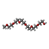
Mass: 356.540 Da / Num. of mol.: 5 / Source method: obtained synthetically / Formula: C21H40O4 / Feature type: SUBJECT OF INVESTIGATION
#3: Chemical ChemComp-P15 / 2,5,8,11,14,17-HEXAOXANONADECAN-19-OL


Mass: 296.357 Da / Num. of mol.: 2 / Source method: obtained synthetically / Formula: C13H28O7 / Feature type: SUBJECT OF INVESTIGATION
#4: Chemical
ChemComp-OLA / OLEIC ACID


Mass: 282.461 Da / Num. of mol.: 12 / Source method: obtained synthetically / Formula: C18H34O2 / Feature type: SUBJECT OF INVESTIGATION
#5: Chemical ChemComp-CLR / CHOLESTEROL


Mass: 386.654 Da / Num. of mol.: 1 / Source method: obtained synthetically / Formula: C27H46O
#6: Water ChemComp-HOH / water


Mass: 18.015 Da / Num. of mol.: 74 / Source method: isolated from a natural source / Formula: H2O

-
Details

Has ligand of interestY
Has protein modificationY

-
Experimental details

-
Experiment

ExperimentMethod: X-RAY DIFFRACTION / Number of used crystals: 1

-
Sample preparation

CrystalDensity Matthews: 2.83 Å3/Da / Density % sol: 56.49 % / Description: orthorhombic crystals
Crystal growTemperature: 293 K / Method: lipidic cubic phase / Details: NaCH3COO, PEG 400, NaCl, PPG P40

-
Data collection

DiffractionMean temperature: 123 K / Serial crystal experiment: N
Diffraction sourceSource: SYNCHROTRON / Site: APS / Beamline: 23-ID-B / Wavelength: 1.0332 Å
DetectorType: DECTRIS EIGER X 16M / Detector: PIXEL / Date: Feb 27, 2020
RadiationProtocol: SINGLE WAVELENGTH / Monochromatic (M) / Laue (L): M / Scattering type: x-ray
Radiation wavelengthWavelength: 1.0332 Å / Relative weight: 1
ReflectionResolution: 2.1→30 Å / Num. obs: 31292 / % possible obs: 96.8 % / Redundancy: 6.9 % / CC1/2: 0.93 / CC star: 0.982 / Rmerge(I) obs: 0.191 / Net I/σ(I): 9.2
Reflection shellResolution: 2.1→2.18 Å / Rmerge(I) obs: 0.638 / Mean I/σ(I) obs: 1.2 / Num. unique obs: 2492 / CC1/2: 0.677 / CC star: 0.898

-
Processing

Software
NameVersionClassification
PHENIX(1.20.1_4487: ???)refinement
HKL-2000data reduction
HKL-2000data scaling
PHASERphasing
RefinementMethod to determine structure: MOLECULAR REPLACEMENT / Resolution: 2.1→29.44 Å / SU ML: 0.22 / Cross valid method: FREE R-VALUE / σ(F): 1.35 / Phase error: 21.44 / Stereochemistry target values: ML
RfactorNum. reflection% reflection
Rfree0.2141 1677 5.41 %
Rwork0.1845 --
obs0.1861 30995 96.03 %
Solvent computationShrinkage radii: 0.9 Å / VDW probe radii: 1.11 Å / Solvent model: FLAT BULK SOLVENT MODEL
Refinement stepCycle: LAST / Resolution: 2.1→29.44 Å
ProteinNucleic acidLigandSolventTotal
Num. atoms2777 0 387 74 3238
Refine LS restraints
Refine-IDTypeDev idealNumber
X-RAY DIFFRACTIONf_bond_d0.0033294
X-RAY DIFFRACTIONf_angle_d0.6954398
X-RAY DIFFRACTIONf_dihedral_angle_d13.7221321
X-RAY DIFFRACTIONf_chiral_restr0.038504
X-RAY DIFFRACTIONf_plane_restr0.004527
LS refinement shell
Resolution (Å)Rfactor RfreeNum. reflection RfreeRfactor RworkNum. reflection RworkRefine-ID% reflection obs (%)
2.1-2.160.28021010.28981886X-RAY DIFFRACTION75
2.16-2.230.25441050.25772220X-RAY DIFFRACTION88
2.23-2.310.24651280.22932416X-RAY DIFFRACTION95
2.31-2.40.28651380.21072463X-RAY DIFFRACTION98
2.4-2.510.24931580.22454X-RAY DIFFRACTION99
2.51-2.650.21831430.17822485X-RAY DIFFRACTION99
2.65-2.810.17061680.16382503X-RAY DIFFRACTION100
2.81-3.030.18711520.17012519X-RAY DIFFRACTION100
3.03-3.330.24851320.18642541X-RAY DIFFRACTION99
3.33-3.810.2311500.17322558X-RAY DIFFRACTION100
3.81-4.80.17721450.15872586X-RAY DIFFRACTION100
4.8-29.440.21761570.19612687X-RAY DIFFRACTION99
Refinement TLS params.

Method: refined / Refine-ID: X-RAY DIFFRACTION

IDL112)L122)L132)L222)L232)L332)S11 (Å °)S12 (Å °)S13 (Å °)S21 (Å °)S22 (Å °)S23 (Å °)S31 (Å °)S32 (Å °)S33 (Å °)T112)T122)T132)T222)T232)T332)Origin x (Å)Origin y (Å)Origin z (Å)
11.64280.15410.01451.3469-0.29110.2843-0.02010.0035-0.02090.0596-0.0578-0.04470.0446-0.0194-0.00010.23640.00810.00360.1863-0.00660.279629.45517.96846.658
20.5176-0.05260.09980.0373-0.45710.69950.01430.2998-0.0498-0.1949-0.02110.0610.2014-0.22020.00010.52430.019-0.03320.5137-0.03190.406714.17516.84621.635
Refinement TLS group
IDRefine-IDRefine TLS-IDSelection detailsAuth asym-IDAuth seq-ID
1X-RAY DIFFRACTION1( CHAIN A AND RESID 70:223 )A70 - 223
2X-RAY DIFFRACTION2( chain 'A' and (resid 224 through 256 or resid 1001 through 1106 or resid 900 or resid 270 through 347 ))A224 - 347

+
About Yorodumi

-
News

-
Feb 9, 2022. New format data for meta-information of EMDB entries

New format data for meta-information of EMDB entries

  • Version 3 of the EMDB header file is now the official format.
  • The previous official version 1.9 will be removed from the archive.

Related info.:EMDB header

External links:wwPDB to switch to version 3 of the EMDB data model

-
Aug 12, 2020. Covid-19 info

Covid-19 info

URL: https://pdbj.org/emnavi/covid19.php

New page: Covid-19 featured information page in EM Navigator.

Related info.:Covid-19 info / Mar 5, 2020. Novel coronavirus structure data

+
Mar 5, 2020. Novel coronavirus structure data

Novel coronavirus structure data

Related info.:Yorodumi Speices / Aug 12, 2020. Covid-19 info

External links:COVID-19 featured content - PDBj / Molecule of the Month (242):Coronavirus Proteases

+
Jan 31, 2019. EMDB accession codes are about to change! (news from PDBe EMDB page)

EMDB accession codes are about to change! (news from PDBe EMDB page)

  • The allocation of 4 digits for EMDB accession codes will soon come to an end. Whilst these codes will remain in use, new EMDB accession codes will include an additional digit and will expand incrementally as the available range of codes is exhausted. The current 4-digit format prefixed with “EMD-” (i.e. EMD-XXXX) will advance to a 5-digit format (i.e. EMD-XXXXX), and so on. It is currently estimated that the 4-digit codes will be depleted around Spring 2019, at which point the 5-digit format will come into force.
  • The EM Navigator/Yorodumi systems omit the EMD- prefix.

Related info.:Q: What is EMD? / ID/Accession-code notation in Yorodumi/EM Navigator

External links:EMDB Accession Codes are Changing Soon! / Contact to PDBj

+
Jul 12, 2017. Major update of PDB

Major update of PDB

  • wwPDB released updated PDB data conforming to the new PDBx/mmCIF dictionary.
  • This is a major update changing the version number from 4 to 5, and with Remediation, in which all the entries are updated.
  • In this update, many items about electron microscopy experimental information are reorganized (e.g. em_software).
  • Now, EM Navigator and Yorodumi are based on the updated data.

External links:wwPDB Remediation / Enriched Model Files Conforming to OneDep Data Standards Now Available in the PDB FTP Archive

-
Yorodumi

Thousand views of thousand structures

  • Yorodumi is a browser for structure data from EMDB, PDB, SASBDB, etc.
  • This page is also the successor to EM Navigator detail page, and also detail information page/front-end page for Omokage search.
  • The word "yorodu" (or yorozu) is an old Japanese word meaning "ten thousand". "mi" (miru) is to see.

Related info.:EMDB / PDB / SASBDB / Comparison of 3 databanks / Yorodumi Search / Aug 31, 2016. New EM Navigator & Yorodumi / Yorodumi Papers / Jmol/JSmol / Function and homology information / Changes in new EM Navigator and Yorodumi

Read more